Announcing wDSSR: The Next-Generation Web Interface to X3DNA-DSSR
Dear 3DNA/DSSR Community,
We are thrilled to announce the official launch of wDSSR (https://web.x3dna-dssr.org/), the powerful new web interface to the X3DNA-DSSR analytical engine.
Developed by Drs. Shuxiang Li and Xiang-Jun Lu and supported by NIH grant R24GM153869, wDSSR represents a major leap forward from our highly popular 2019 Web 3DNA 2.0 framework. While Web 3DNA 2.0 has faithfully served the community for the analysis, visualization, and modeling of 3D nucleic acid structures, wDSSR was built from the ground up to take full advantage of modern web technologies and the latest DSSR backend capabilities.
A Modern, Streamlined Scientific Workflow
We have completely overhauled the user interface to provide a clean, intuitive, and task-driven experience. The core modeling and analysis tools are now seamlessly organized into a logical, single-word scientific workflow: Analyze, Rebuild, Model, Circularize, Mutate, Assemble, and Visualize.
Spotlight Feature: The "Assemble" Module
One of the most exciting upgrades is the newly renamed Assemble tab (formerly "Composite"). This advanced composite model builder allows you to effortlessly construct complex, higher-order models by linking any combination of nucleic acid duplexes or protein-DNA/RNA complexes. You can quickly connect up to six distinct target structures, ranging from simple linked A-DNA and B-DNA duplexes to large, protein-decorated structural assemblies.
Immediate Global Adoption
Although wDSSR has just launched, we are incredibly humbled to share that it is already seeing rapid worldwide adoption! According to recent network infrastructure data, the new interface is actively being used by researchers across North America, South America, Europe, and Asia. Within just a few days, we have recorded active sessions from prestigious institutions around the globe, including:
- The Weizmann Institute of Science in Israel
- Katholieke Universiteit Leuven in Belgium
- Queen's University in Canada
- Universidad Nacional Autonoma de Mexico (UNAM) in Mexico
- Emory University and the Wadsworth Centers Laboratories and Research in the United States
- Jawaharlal Nehru University and the China Education and Research Network in Asia
How to Cite
While a dedicated paper for wDSSR is currently in preparation, researchers should cite the server using its URL (https://web.x3dna-dssr.org/) alongside the 2019 Web 3DNA 2.0 paper and the foundational 2015 DSSR paper. Full details and funding acknowledgements can be found on our newly consolidated About page.
We invite you all to try out the new wDSSR platform! As always, your feedback is invaluable to us, and we encourage you to share your thoughts, questions, and structural models via the newly updated Questions & Feedback link in the wDSSR footer.
Happy modeling!
The blocview script in 3DNA has been created as a handy tool to effectively reveal key features of small to medium-sized nucleic acid structures. Specifically, the bloc part of the name means ‘block’, i.e., the rectangular block in Calladine-Drew style schematic representation to distinguish bases by size (larger purine vs. smaller pyrimidine), identity (red for A, yellow for C, green for G, and blue for T), and groove (minor edge in black). The view part stands for the most extended view, as defined by the principal axes of inertia. Implementation-wise, blocview calls several 3DNA utility programs and MolScript (for protein ribbons and nucleic acid backbone rods) to prepare the scenes, and then uses Raster3D (specifically, render) or PyMol to generate a PNG image.
The blocview script was originally written in Perl. As of 3DNA v2.1, I decided to switch the scripting language to Ruby for its consistent object-oriented style, succinct and flexible syntax. Previously available Perl scripts are now moved out of the default 3DNA executable directory $X3DNA/bin/ into $X3DNA/perl_scripts/. The blocview script has been re-written in Ruby and set as the default (at $X3DNA/bin/blocview); the original Perl version is renamed blocview.pl (at $X3DNA/perl_scripts/blocview.pl) to avoid confusion. The command line help message, available via blocview -h, is as below:
------------------------------------------------------------------------
Generate a schematic image which combines base block representation
with protein ribbon. The image has informative color coding for the
nucleic acid part and is set in the "best view" by default. Raster3D
(or PyMOL) and ImageMagick must be installed.

Usage:
        blocview [options] PDBFile
Examples:
        blocview -i 355d.png 355d.pdb
             # generate image '355d.png'; display 355d.png
Options:
------------------------------------------------------------------------
     --imgfile, -i <s>:   name of image file (default: blocview.png)
     --r3dfile, -r <s>:   name of .r3d file (default: blocview.r3d)
   --dpi-pymol, -d <i>:   create PyMOL ray-traced image at specific DPI
       --scale, -s <f>:   set scale factor (for 'render' of Raster3D)
        --xrot, -x <f>:   rotation angle about x-axis
        --yrot, -y <f>:   rotation angle about y-axis
        --zrot, -z <f>:   rotation angle about z-axis
        --original, -o:   use original coordinates
  --ball-and-stick, -b:   get a ball-and-stick image
     --p-base-ring, -c:   use only P and base ring atoms
           --no-ds, -n:   do not show double-helix ribbon
         --protein, -p:   set best view based on protein atoms
             --all, -a:   set best view based on all atoms
         --version, -v:   Print version and exit
            --help, -h:   Show this message
Using the x-ray crystal structure of d(GGCCAATTGG) complexed with netropsin (1z8v) in the minor groove as an example, the command to run is as follows:
blocview -i 1z8v.png 1z8v.pdb
# The following two forms are also fine
#     blocview --imgfile 1z8v.png 1z8v.pdb
#     blocview --imgfile=1z8v.png 1z8v.pdb
# The Perl version can be run like this:
#     $X3DNA/perl_scripts/blocview.pl -i=1z8v.png 1z8v.pdb
The image, named 1z8v.png, is shown below. Note that it is generated automatically from the PDB-formatted data file 1z8v.pdb. In this representation, one can see clearly that there are two unpaired Gs (green block) at each 5′-end of the two DNA chains (red and yellow rods), and a drug molecule (ball-and-stick) binds in the minor groove (black edge of the rectangular blocks). Moreover, the deformation in propeller and buckle is obvious in this schematic presentation.

Over the years, blocview-generated images have been used in NDB for virtually all nucleic acid structures (see for example, the NDB atlas gallery for x-ray drug-DNA complexes). It’s worth noting that such simple images have also be adopted by the RCSB PDB, prominently at the summary page, for nucleic acid containing structures (see PDB entry 1z8v). Given the effectiveness of blocview-generated schematic representation and its adoption by the NDB and PDB, I’m hopeful that blocview will be more widely used by the general DNA/RNA structure community. As always, I value user’s feedback in continuously refining the script.

One of 3DNA’s unique features is the simplified rectangular block representation of bases and base-pairs, as shown in the figure below. This type of schematic depiction was first made popular by Calladine and Drew (see their book titled Understanding DNA — The Molecule & How It Works), thus I usually call it the Calladine-Drew style representation.

By default, a base-pair [BP, (a)] has dimensions of 10×4.5×0.5 (Å); a purine [R, (b) left] 4.5×4.5×0.5 (Å); a pyrimidine [Y, (b) right] 3×4.5×0.5 (Å); and a mean base [M, (c)], which is exactly half of the base-pair, 5×4.5×0.5 (Å).
The blocks are stored into separate files: Block_BP.alc, Block_R.alc, and Block_Y.alc for BP, R and Y respectively. To use M for R and Y (i.e., set R and Y to be of equal size), simple copy file Block_M.alc to overwrite Block_R.alc and Block_Y.alc in the current working directory for local effect, or the 3DNA installation directory ($X3DNA/config/) for global impact. These blocks are used in the rebuilding and visualization components of 3DNA.
Following SCHNArP, 3DNA uses alchemy, a simple chemical file format, to specify explicitly the nodes (atoms) and edges (bonds) of a rectangular block. Three file formats (alchemy, MDL molfile, and Tripos mol2), supported by RasMol v2.6 (the most popular molecular graphics visualization program in the 1990s), serve the purpose of specifying the rectangular block. I cannot recall exactly why I picked up -alchemy instead of -mdl and -mol2, perhaps because of its simplicity: I played around with sample alchemy files and came up with the alchemy rectangular block files used by SCHNArP, without much difficulty.
As an example, Block_BP.alc has the following content:
   12 ATOMS,    12 BONDS
    1 N      -2.2500   5.0000   0.2500
    2 N      -2.2500  -5.0000   0.2500
    3 N      -2.2500  -5.0000  -0.2500
    4 N      -2.2500   5.0000  -0.2500
    5 C       2.2500   5.0000   0.2500
    6 C       2.2500  -5.0000   0.2500
    7 C       2.2500  -5.0000  -0.2500
    8 C       2.2500   5.0000  -0.2500
    9 C      -2.2500   5.0000   0.2500
   10 C      -2.2500  -5.0000   0.2500
   11 C      -2.2500  -5.0000  -0.2500
   12 C      -2.2500   5.0000  -0.2500
    1     1     2
    2     2     3
    3     3     4
    4     4     1
    5     5     6
    6     6     7
    7     7     8
    8     5     8
    9     9     5
   10    10     6
   11    11     7
   12    12     8
Observant viewers may notice that nodes 1-4 are specified as nitrogens (N) which have exactly the same coordinates as 9-12 (carbons, C). This is a little trick to make RasMol display the minor groove edge in a different color (blue for N) than the other five sides of the rectangular (gray for C), as shown in the following figure:

Note that the rectangular is preset in the standard base reference frame. Thus the nodes have y-coordinates of +5 Å and -5 Å along the long edge of the base pair, and x-coordinates of +2.25 Å and -2.25 Å along the short edge.
As an extra bonus of storing the rectangular blocks in external alchemy text files, the dimensions of the blocks can be readily changed. For example, the thickness of a block (z-coordinates) can be easily increased from 0.5 to 1.0 Å to make it thicker. Moreover, the blocks do not need to be rectangular either — they can appear to be triangular blocks.
It’s worth noting that while extensively used in 3DNA for schematic representations, the alchemy format has largely become a legacy in cheminformatics/bioinformatics nowadays. Searching the internet, I cannot find the specification of the format. Moreover, the support of alchemy is quite limited and buggy in molecular graphics visualization programs most widely used today: PyMOL does not understand this format at all; RasMol v2.7 has a bug in interpreting it; only Jmol can properly read 3DNA base-pair rectangular block files in alchemy [see initial discussion and follow-up]. To resolve the issues associated with alchemy format, and thus to make 3DNA base-pair block schematics more widely available, I have recently added a converter in v2.1 to readily transform alchemy to MDL molfile, a format consistently supported by PyMOL, Jmol and RasMol. I’ll talk about this feature in another post.
The conformation of the five-membered sugar ring in DNA/RNA structures can be characterized by the five consecutive endocyclic torsion angles (see Figure below), i.e.,
ν0: C4′-O4′-C1′-C2′
ν1: O4′-C1′-C2′-C3′
ν2: C1′-C2′-C3′-C4′
ν3: C2′-C3′-C4′-O4′
ν4: C3′-C4′-O4′-C1′

Due to the ring constraint, the conformation can be characterized approximately by 5-3=2 parameters. Using the concept of pseudorotation of the sugar ring, the two parameters are the amplitude (τm) and phase angle (P).
One set of widely used formula to convert the five torsion angles to the pseudorotation parameters is due to Altona & Sundaralingam (1972): Conformational Analysis of the Sugar Ring in Nucleosides and Nucleotides. A New Description Using the Concept of Pseudorotation [J. Am. Chem. Soc., 94(23), pp. 8205–8212]. The concept is easily illustrated with an example — here with the G4 sugar ring on chain A of the Dickerson dodecamer (1bna), using Octave/Matlab code:
# xyz coordinates of the G4 sugar ring on chain A of 1bna
# ATOM     63  C4'  DG A   4      21.393  16.960  18.505  1.00 53.00
# ATOM     64  O4'  DG A   4      20.353  17.952  18.496  1.00 38.79
# ATOM     65  C3'  DG A   4      21.264  16.229  17.176  1.00 56.72
# ATOM     67  C2'  DG A   4      20.793  17.368  16.288  1.00 40.81
# ATOM     68  C1'  DG A   4      19.716  17.901  17.218  1.00 30.52

# endocyclic torsion angles:
v0 = -26.7; v1 = 46.3; v2 = -47.1; v3 = 33.4; v4 = -4.4;
Pconst = sin(pi/5) + sin(pi/2.5);  # 1.5388
P0 = atan2(v4 + v1 - v3 - v0, 2.0 * v2 * Pconst); # 2.9034
tm = v2 / cos(P0)  # amplitude: 48.469
P = 180/pi * P0  # phase angle: 166.35 [P + 360 if P0 < 0]
The Altona & Sundaralingam (1972) pseudorotation parameters are what have been adopted in 3DNA. The Curves+ program, on the other hand, uses another set of formula due to Westhof & Sundaralingam (1983): A Method for the Analysis of Puckering Disorder in Five-Membered Rings: The Relative Mobilities of Furanose and Proline Rings and Their Effects on Polynucleotide and Polypeptide Backbone Flexibility. [J. Am. Chem. Soc., 105(4), pp. 970–976]. The two sets of formula — Altona & Sundaralingam (1972) and Westhof & Sundaralingam (1983) — give slightly different numerical values for the two pseudorotation parameters (see below).
Since Curves+ and 3DNA are currently the most commonly used programs for conformational analysis of nucleic acid structures, the subtle differences in these two pseudorotation parameters may cause confusions for users who use both programs. With the same G4 (on chain A of 1bna) sugar ring, here is the Octave/Matlab script showing how Curve+ calculates the pseudorotation parameters:
# xyz coordinates of the G4 sugar ring on chain A of 1bna
# endocyclic torsion angles, same as above
v0 = -26.7; v1 = 46.3; v2 = -47.1; v3 = 33.4; v4 = -4.4;

v = [v2, v3, v4, v0, v1]; # reorder them into vector v[]
A = 0; B = 0;
for i = 1:5
    t = 0.8 * pi * (i - 1);
    A += v(i) * cos(t);
    B += v(i) * sin(t);
end
A *= 0.4;   # -48.476
B *= -0.4;  # 11.516

tm = sqrt(A * A + B * B);  # 49.825

c = A/tm; s = B/tm;
P = atan2(s, c) * 180 / pi;  # 166.64
For this specific example, i.e., the G4 sugar ring on chain A of 1bna, the pseudorotation parameters as calculated by 3DNA following Altona & Sundaralingam (1972) and Curves+ following Westhof & Sundaralingam (1983) are as follows:
|
amplitude (τm) |
phase angle (P) |
| 3DNA |
48.469 |
166.35 |
| Curves+ |
49.825 |
166.64 |
Needless to say, for the majority of cases like the one shown here, the differences are subtle; very few people would notice them or be bothered at all. For those who do care about such little details, however, this post shows where the discrepancies really come from.
In the field of nucleic acid structural analysis, it seems fair to say that Curves+ and 3DNA are nowadays the top two choices. To the best of my knowledge, these two programs are also the only ones that confirm to the standard base reference frame. Moreover, as noted in my previous post, Curves+ and 3DNA are “constructive competitors” with complementary functionality: Curves+ is unique in providing a curvilinear helical axis, a bending analysis, a full description of groove widths and depths and its seamless integration to the analysis of molecular dynamics trajectories, while 3DNA’s strength lies in its cohesrent approach combining analysis, rebuilding, and visualization into one package.
Given the complementarity between Curve+ and 3DNA, it makes sense to build a ‘bridge’ between the two so users can easily take advantage of both programs. Starting from 3DNA v1.5, find_pair has the -c option to generate input for Curves directly from a PDB file. Over the years, this option appears to have received little attention — at least, I am not aware of any literature reference to it. Now, the updated Curves+ program has introduced the new lib name list variable, among other changes. I have thus added the -curves+ option (abbreviation -c+) to find_pair to make its output compatible with Curves+.
As always, the point/process is best illustrated with an example — here with the Dickerson B-DNA dodecamer solved at high resolution by Williams et al. (PDB entry 355d).
find_pair -c+ 355d.pdb 355d-curves+.inp
The generated file 355d-curves+.inp has the following content:
&inp file=355d.pdb,
     lis=355d,
     fit=.t.,
     lib=/Users/xiangjun/Curves+/standard,
     isym=1,
&end
    2    1   -1    0    0
    1    2    3    4    5    6    7    8    9   10   11   12
   24   23   22   21   20   19   18   17   16   15   14   13
which can be fed into Curves+ as below,
Cur+ < 355d-curves+.inp
The four output files are: 355d.cda, 355d.lis, 355d_X.pdb and 355d_b.pdb.
Please note the followings:
- The environment variable CURVES_PLUS_STDLIB should be set, pointing to the directory where Curves+ is installed (containing files
standard_b.lib and standard_s.lib). In the example above, CURVES_PLUS_STDLIB is set to /Users/xiangjun/Curves+.
- The
find_pair -c+ option (currently) is applicable only to double helical DNA/RNA structures, the most common application scenario.
- The
-c+ option ignores HETATM records, in accordance with Curves+ where proteins, water and HETATM are automatically removed at input. (see Curves+ user manual, section Input data)
- To run
Cur+ < 355d-curves+.inp again in the same folder, the four output files must first be deleted (e.g., rm -f 355d.cda 355d.lis 355d_[Xb].pdb). This is best taken care of via a script.
Obviously, the nucleic acid structure community benefits the most to have both Curves+ and 3DNA at its disposal and be able to easily switch between them — hopefully, the find_pair -c+ option would serve as such a ‘bridge’.
From the very beginning, 3DNA calculates a set of nucleic acid backbone parameters, including the six main chain torsion angles (α, β, γ, δ, ε, and ζ) around the covalent bonds, χ about the glycosidic bond, and the sugar pucker (see figure below). For double helical structures, the standard analyze output (.out file) has a section for “Main chain and chi torsion angles,” and another dedicated to “Sugar conformational parameters”. Based on my experience/understanding, these two parts are well recognized and utlizied by 3DNA users. What has receive little attention (in spite of the several posts I’ve written on the topic), though, is 3DNA’s applicability to single-stranded (ss) RNA structures for the backbone torsions, among other parameters. Using the fully refined crystal structure of the Haloarcula marismortui large ribosomal subunit (PDB entry 1jj2) as an example, the procedure is below:
find_pair -s 1jj2.pdb 1jj2.nts
analyze 1jj2.nts
    # or the above two steps can be combined:
find_pair -s 1jj2.pdb stdout | analyze stdin
    # see output file '1jj2.outs'
In retrospect, the fact that 3DNA has been little used for RNA backbone conformational analysis is of no surprise:
- While base-pair parameters have different (oftentimes confusing) definitions, these backbone parameters are pretty “standard” — thus, for example, any program for DNA/RNA structural analysis would give the same numerical values for α or χ torsion angles.
- The two-step process as illustrated above is a bit awkward, and the torsions are “buried” among many other parameters.
- 3DNA is more directly “linked” (conceivably) to DNA base pairs than to RNA backbone.
So while adapting the Zp parameter for ss DNA/RNA structures in 3DNA v2.1, I also take this opportunity to add the -torsion option to analyze with the following handy features:
- Streamline the calculation by starting directly from a PDB file and output only backbone parameters. So the above example can be shortened to
analyze -t=1jj2.tor 1jj2.pdb; the output file is named 1jj2.tor.
- Classify backbone into BI/BII conformation, and base χ into syn / anti.
- Add pseudo-torsions, and Zp and Dp as defined by Richardson et al.
- Handle pseudouridine sensibly, and work also for nucleic acid structure with only backbone atoms.
- Be easy to use, efficient and robust — it takes ~1 second to process the large ribosomal subunit 1jj2 (with 2876 nucleotides consisting of 23S rRNA and 5S rRNA) on my MacBook Air.
Overall, analyze -torsion is designed to be pragmatic and allows for automatic processing of all NDB entries or molecular dynamics trajectories. Given below is an excerpt of the three sections from an analyze -torsion run on 1jj2:
****************************************************************************
Main chain and chi torsion angles:

Note: alpha:   O3'(i-1)-P-O5'-C5'
      beta:    P-O5'-C5'-C4'
      gamma:   O5'-C5'-C4'-C3'
      delta:   C5'-C4'-C3'-O3'
      epsilon: C4'-C3'-O3'-P(i+1)
      zeta:    C3'-O3'-P(i+1)-O5'(i+1)

      chi for pyrimidines(Y): O4'-C1'-N1-C2
          chi for purines(R): O4'-C1'-N9-C4

          If chi is in range [-90, +90], syn conformation
              otherwise, it is in anti conformation

          e-z: epsilon - zeta
              BI:  e-z = [-160, +20]
              BII: e-z = [+20, +200]

              base      chi         alpha    beta   gamma   delta  epsilon   zeta     e-z
   1 0:..10_:[..U]U   -62.5(syn)     ---     ---     56.2    74.0   142.2   -87.8  -130.1(BI)
   2 0:..11_:[..A]A   171.5(anti)   173.2  -161.0   168.5    84.0  -112.1   -65.4   -46.7(BI)
   3 0:..12_:[..U]U  -167.7(anti)   -70.7   168.4    53.0    78.5  -128.5   -46.4   -82.1(BI)
   4 0:..13_:[..G]G  -172.5(anti)   -61.8   170.4    67.7    73.5  -166.7   -79.6   -87.1(BI)
   5 0:..14_:[..C]C  -166.0(anti)   -73.0  -172.5    55.1    83.2  -143.3   -77.7   -65.6(BI)
   6 0:..15_:[..C]C  -155.5(anti)   -60.9   174.1    47.3    80.3  -154.4   -71.2   -83.2(BI)

****************************************************************************
Pseudo (virtual) eta/theta torsion angles:

Note: eta:    C4'(i-1)-P(i)-C4'(i)-P(i+1)
      theta:  P(i)-C4'(i)-P(i+1)-C4'(i+1)

      eta':   C1'(i-1)-P(i)-C1'(i)-P(i+1)
      theta': P(i)-C1'(i)-P(i+1)-C1'(i+1)

      eta":   Borg(i-1)-P(i)-Borg(i)-P(i+1)
      theta": P(i)-Borg(i)-P(i+1)-Borg(i+1)

              base      eta   theta    eta'  theta'    eta"  theta"
   1 0:..10_:[..U]U    ---     ---     ---     ---     ---     ---
   2 0:..11_:[..A]A  -174.6  -129.7   177.0  -127.7  -157.5   -75.5
   3 0:..12_:[..U]U   149.1  -105.1   174.1  -101.2  -111.0   -69.4
   4 0:..13_:[..G]G   169.0  -172.5  -156.6  -169.2   -93.3  -137.1
   5 0:..14_:[..C]C   176.2  -143.4   179.6  -140.6  -144.6  -120.6
   6 0:..15_:[..C]C   165.0  -147.7   177.4  -146.8  -149.2  -121.7

****************************************************************************
Sugar conformational parameters:

Note: v0: C4'-O4'-C1'-C2'
      v1: O4'-C1'-C2'-C3'
      v2: C1'-C2'-C3'-C4'
      v3: C2'-C3'-C4'-O4'
      v4: C3'-C4'-O4'-C1'

      tm: the amplitude of pucker
      P:  the phase angle of pseudorotation

      Zp: z-coordinate of the 3' phosphorus atom (P) expressed in the
            standard base reference frame; it's POSITIVE when P is on
            the +z-axis side (base in anti conformation); NEGATIVE if
            P is on the -z-axis side (base in syn conformation)
      Dp: perpendicular distance of the 3' P atom to the glycosydic bond
            [as per the MolProbity paper of Richardson et al. (2010)]

              base       v0      v1      v2      v3      v4     tm       P    Puckering    Zp      Dp
   1 0:..10_:[..U]U   -11.3   -15.4    34.5   -41.8    33.5    41.6    33.8    C3'-endo   -0.13    3.53
   2 0:..11_:[..A]A    11.4   -30.2    36.9   -31.5    12.6    36.9     1.2    C3'-endo    4.74    4.78
   3 0:..12_:[..U]U     3.6   -29.3    42.4   -41.3    23.8    43.6    13.9    C3'-endo    4.67    4.82
   4 0:..13_:[..G]G   -13.0   -17.8    39.8   -47.9    38.5    47.8    33.7    C3'-endo    4.45    4.46
   5 0:..14_:[..C]C     6.0   -28.4    38.9   -36.5    19.2    39.5    10.1    C3'-endo    4.57    4.70
   6 0:..15_:[..C]C     1.9   -26.4    39.6   -39.6    23.7    41.2    16.0    C3'-endo    4.32    4.61
Given the x-, y-, and z-coordinates of four points (a-b-c-d) in 3-dimensional (3D) space, how to calculate the torsion angle? Overall, this is a well-solved problem in structural biology and chemistry; one can find a description of torsion angle in many text books and on-line documents. The algorithm for its calculation is implementated in virtually every software package in computational structural biology and chemistry.
As basic as the concept is, however, it is important (based on my experience) to have a clear understanding of how torsion angle is defined in order to really get into the 3D world. Here is a worked example using Octave/Matlab of my simplified, geometry-based implementation of calculating torsion angle, including how to determine its sign. No theory or (complicated) mathematical formula, just a step-by-step illustration of how I solve this problem.
- Coordinates of four points are given in variable
abcd:
abcd = [ 21.350  31.325  22.681
         22.409  31.286  21.483
         22.840  29.751  21.498
         23.543  29.175  22.594 ];
- Two auxiliary functions: norm_vec() to normalize a vector; get_orth_norm_vec() to get the orthogonal component (normalized) of a vector with reference to another vector, which should have already been normalized.
function ovec = norm_vec(vec)
    ovec = vec / norm(vec);
endfunction
function ovec = get_orth_norm_vec(vec, vref)
    temp = vec - vref * dot(vec, vref);
    ovec = norm_vec(temp);
endfunction
- Get three vectors: b_c is the normalized vector b→c; b_a_orth is the orthogonal component (normalized) of vector b→a with reference to b→c; c_d_orth is similarly defined, as the orthogonal component (normalized) of vector c→d with reference to b→c.
b_c = norm_vec(abcd(3, :) - abcd(2, :))
    % [0.2703158  -0.9627257   0.0094077]
b_a_orth = get_orth_norm_vec(abcd(1, :) - abcd(2, :), b_c)
    % [-0.62126  -0.16696   0.76561]
c_d_orth = get_orth_norm_vec(abcd(4, :) - abcd(3, :), b_c)
    % [0.41330   0.12486   0.90199]
- Now the torsion angle is defined as the angle between the two vectors, b_a_orth and c_d_orth, and can be easily calculated by their dot product. The sign of the torsion angle is determined by the relative orientation of the cross product of the same two vectors with reference to the middle vector b→c. Here they are in opposite direction, thus the torsion angle is negative.
angle_deg = acos(dot(b_a_orth, c_d_orth)) * 180 / pi  % 65.609
sign = dot(cross(b_a_orth, c_d_orth), b_c)  % -0.91075
if (sign < 0)
    ang_deg = -angle_deg  % -65.609
endif
A related concept is the so-called dihedral angle, or more generally the angle between two planes. As long as the normal vectors to the two corresponding planes are defined, the angle between them is easy to work out.
It’s worth noting that the helical twist angle in SCHNAaP and 3DNA is calculated similarly.
Backbone conformation of nucleic acid structures is most characterized by a set of 6 torsion angles (α, β, γ, δ, ε, and ζ) around the consecutive chemical bonds, chi (χ) quantifying the relative base/sugar orientation, plus the sugar pucker.

This large number of DNA/RNA backbone conformational parameters is in striking contrast to the two torsion angles (φ and ψ) in protein structures, routinely employed in Ramachandran plot. Over the years, the nucleic acid community has come up with simplified ways to represent DNA/RNA backbone conformation. Thus far, the most widely used one is the pseudo-torsion angles (See figure below) η: C4′(i-1)-P(i)-C4′(i)-P(i+1) and θ: P(i)-C4′(i)-P(i+1)-C4′(i+1).

The history of the P—C4′ virtual-bond concept and its application in RNA structure analysis have recently been reviewed by Pyle et al. in A new way to see RNA [Q Rev Biophys. 2011, 44(4), 433—466], where the following three contributions are highlighted:
- Olson (1980). Configurational statistics of polynucleotide chains. An updated virtual bond model to treat effects of base stacking., Macromolecules 13(3), 721—728.
- Malathi & Yathindra (1980). A novel virtual bond scheme to probe ordered and random coil conformations of nucleic acids: Configurational statistics of polynucleotide chains. Current Science, 49, 803—807.
- Duarte & Pyle (1998). Stepping through an RNA structure: A novel approach to conformational analysis. Journal of Molecular Biology, 284, 1465—1478.
More recently, Pyle et al. also employed a modified version of the pseudo-torsions, η′: C1′(i-1)-P(i)-C1′(i)-P(i+1) and θ′: P(i)-C1′(i)-P(i+1)-C1′(i+1), i.e., using C1′ instead of C4′, and found that:
The η′ and θ′ torsions are more suitable when interpreting crystallographic density because the C1′ atom is covalently bound to the nucleoside base and therefore can be more easily and accurately located within a low-resolution map.
While implementing the -torsion option to analyze to make it more explicit that 3DNA readily calculates conventional backbone torsion angles, I also take this opportunity to add the pseudo-torsion angles — η/θ and η′/θ′, among other new parameters. Moreover, while I am at it, I cannot help but also compute yet another set of pseudo-torsion angles: η″/θ″. Here, instead of C1′ or C4′, the origin of the base reference frame is employed; it can be taken as a _pseudo_-atom more accurately defined by the base plane than any real single atom.
The usefulness of η″/θ″, especially in comparison with η/θ and η′/θ′, remains to be determined. However, only η″/θ″ uniquely takes advantage of the two most accurately determined entities in a nucleic acid structure, the heavy phosphorus atom and the rigid base plane [see discussion (p.16) in the Richardson et al. MolProbity paper, Acta Cryst. (2010). D66, 12–21] Presumably, η″/θ″ provides a new perspective in RNA structural analysis by combining the backbone and the base.
Here is the pseudo-torsions for the yeast phenylalanine transfer RNA (6tna by simply running analyze -torsion=6tna.tor 6tna.pdb):
Pseudo (virtual) eta/theta torsion angles:

Note: eta:    C4'(i-1)-P(i)-C4'(i)-P(i+1)
      theta:  P(i)-C4'(i)-P(i+1)-C4'(i+1)

      eta':   C1'(i-1)-P(i)-C1'(i)-P(i+1)
      theta': P(i)-C1'(i)-P(i+1)-C1'(i+1)

      eta":   Borg(i-1)-P(i)-Borg(i)-P(i+1)
      theta": P(i)-Borg(i)-P(i+1)-Borg(i+1)

              base      eta   theta    eta'  theta'    eta"  theta"
   1 A:...1_:[..G]G    ---   -126.6    ---   -141.5    ---   -130.4
   2 A:...2_:[..C]C   167.8  -168.3   174.6  -152.5  -151.4  -115.4
   3 A:...3_:[..G]G   160.4  -119.8  -171.9  -138.9  -123.6  -119.2
   4 A:...4_:[..G]G   148.0  -164.2   162.1  -159.2  -154.4  -124.6
   5 A:...5_:[..A]A   168.7  -137.6  -175.9  -137.8  -129.5  -115.0
   6 A:...6_:[..U]U   171.8  -145.7  -172.5  -140.5  -131.3  -124.7
   7 A:...7_:[..U]U  -151.0   -47.8  -136.0   -58.6  -117.7   -30.2
   8 A:...8_:[..U]U   160.9   159.7  -161.0  -163.6  -144.2   178.0
   9 A:...9_:[..A]A  -137.0   -48.6  -158.1  -108.9   161.5  -104.7
  10 A:..10_:[2MG]g    33.1  -135.8    93.4  -134.6   134.1  -113.0
  11 A:..11_:[..C]C   167.2  -138.3  -179.4  -137.7  -142.4  -118.7
  12 A:..12_:[..U]U   165.5  -120.7  -179.3  -128.0  -145.8  -106.7
  13 A:..13_:[..C]C   174.1  -173.6  -165.5   179.6  -120.9  -180.0
  14 A:..14_:[..A]A   173.0  -144.0   172.7  -132.4   177.6   -72.7
  15 A:..15_:[..G]G   154.7   110.6  -176.2    85.5   -97.7   -76.9
  16 A:..16_:[H2U]u    76.3    94.1    65.3   119.7  -152.8  -123.8
  17 A:..17_:[H2U]u   -36.7   -79.6   -50.7  -136.6  -142.7  -159.0
  18 A:..18_:[..G]G    -9.7  -166.8    41.7  -158.6    28.9  -120.4
  19 A:..19_:[..G]G  -131.6   -35.8  -122.9   -67.8  -104.3   -10.5
  20 A:..20_:[..G]G   160.9   -93.2  -161.6   -98.9  -174.1  -112.3
  21 A:..21_:[..A]A   -83.6   152.5   -72.8   155.7   -59.1   155.4
  22 A:..22_:[..G]G   164.1   169.4   160.0  -178.5   159.1  -157.6
  23 A:..23_:[..A]A   177.6  -148.5  -174.5  -142.7  -154.5  -114.3
  24 A:..24_:[..G]G   167.2   -98.9  -171.7  -128.6  -127.6   -99.1
  25 A:..25_:[..C]C   151.6  -153.5   167.3  -140.8  -137.7   -84.8
  26 A:..26_:[M2G]g   156.2  -137.4  -175.2  -135.2  -100.0  -104.2
  27 A:..27_:[..C]C   166.2  -145.5  -177.9  -140.4  -129.1  -116.8
  28 A:..28_:[..C]C   164.7  -140.5   175.8  -145.3  -152.7  -123.4
  29 A:..29_:[..A]A   161.2  -145.3   175.7  -144.9  -142.0  -126.0
  30 A:..30_:[..G]G  -173.5  -120.3  -158.4  -133.2  -126.6   -94.4
  31 A:..31_:[..A]A   169.8  -153.1   177.7  -140.4  -124.5   -81.5
  32 A:..32_:[OMC]c   154.4  -126.8  -178.7  -131.3  -104.1  -128.0
  33 A:..33_:[..U]U   170.0  -103.9  -179.9  -152.7  -164.6   143.6
  34 A:..34_:[OMG]g    -4.7  -123.7    41.8  -124.8    31.6   -99.6
  35 A:..35_:[..A]A   163.5  -104.3   176.9  -127.9  -137.5  -128.2
  36 A:..36_:[..A]A   175.9   173.6   180.0  -167.7  -156.4  -118.3
  37 A:..37_:[.YG]g   166.8  -131.7  -174.5  -133.0  -115.1   -82.9
  38 A:..38_:[..A]A   167.7  -121.6  -175.7  -114.3  -109.9   -79.9
  39 A:..39_:[PSU]P   168.3  -146.8  -160.2  -146.4   -98.6  -116.5
  40 A:..40_:[5MC]c   160.6  -138.7   174.0  -141.8  -139.7  -126.5
  41 A:..41_:[..U]U   164.8  -161.4   175.9  -152.3  -150.5  -117.6
  42 A:..42_:[..G]G   174.3  -140.9  -170.3  -145.4  -129.1  -121.3
  43 A:..43_:[..G]G   169.6  -159.0  -176.2  -154.9  -133.7  -133.1
  44 A:..44_:[..A]A   174.0  -121.5  -174.2  -122.0  -143.1   -74.9
  45 A:..45_:[..G]G   174.4  -132.5  -166.2  -128.1  -101.8  -128.9
  46 A:..46_:[7MG]g  -112.8  -113.4  -127.2  -138.3  -139.8  -152.1
  47 A:..47_:[..U]U   -63.2   -53.8    -1.1   -92.0    22.8  -124.7
  48 A:..48_:[..C]C   -84.7    59.6   -20.1     8.9    19.3  -104.5
  49 A:..49_:[5MC]c   -56.8  -140.1   -29.9  -143.6    98.1  -125.4
  50 A:..50_:[..U]U   173.6  -146.4  -178.3  -140.6  -147.6  -117.8
  51 A:..51_:[..G]G   160.8  -148.1  -178.6  -150.7  -140.7  -121.9
  52 A:..52_:[..U]U   164.9  -144.0   175.8  -143.5  -139.9  -114.3
  53 A:..53_:[..G]G   168.2  -140.9  -171.1  -144.0  -121.6  -117.3
  54 A:..54_:[5MU]u   167.0  -131.1   178.3  -124.9  -139.9   -77.0
  55 A:..55_:[PSU]P   167.6  -114.2  -172.8  -155.6  -113.0   146.0
  56 A:..56_:[..C]C    35.0  -121.5    52.6  -126.2    26.5   -83.8
  57 A:..57_:[..G]G   168.4  -148.1  -177.1  -131.1  -115.4  -111.7
  58 A:..58_:[1MA]a  -136.3  -133.3  -106.5  -176.7  -105.3   149.6
  59 A:..59_:[..U]U    23.0  -130.9    33.0  -115.4    48.2   -68.2
  60 A:..60_:[..C]C  -163.6   -54.3  -123.2   -76.4   -79.6   -36.4
  61 A:..61_:[..C]C   125.5  -153.3   169.7  -144.7  -153.8  -123.4
  62 A:..62_:[..A]A   172.5  -139.3  -177.0  -137.6  -150.7  -114.6
  63 A:..63_:[..C]C   165.8  -146.6  -178.5  -149.8  -139.2  -127.8
  64 A:..64_:[..A]A   164.7  -144.9   176.5  -145.8  -145.3  -118.1
  65 A:..65_:[..G]G   170.4  -152.3  -175.5  -151.5  -132.3  -122.1
  66 A:..66_:[..A]A   168.0  -152.0  -177.4  -150.2  -133.0  -118.7
  67 A:..67_:[..A]A   170.9  -141.8  -178.4  -140.4  -134.8  -123.1
  68 A:..68_:[..U]U   164.8  -135.1  -178.9  -137.9  -143.7   -95.2
  69 A:..69_:[..U]U   168.2  -154.9  -174.3  -157.1  -112.2  -144.8
  70 A:..70_:[..C]C   160.6  -153.2   170.7  -153.5  -164.4  -125.1
  71 A:..71_:[..G]G   161.8  -144.3   172.1  -143.1  -145.7  -124.2
  72 A:..72_:[..C]C   176.7  -136.4  -169.3  -134.5  -134.9   -87.1
  73 A:..73_:[..A]A   160.6  -142.8  -179.7  -139.7  -112.8  -104.4
  74 A:..74_:[..C]C  -176.9  -115.9  -163.1  -115.4  -117.2   -68.7
  75 A:..75_:[..C]C   169.8    80.9  -170.0    74.9  -108.5   -91.3
  76 A:..76_:[..A]A    ---     ---     ---     ---     ---     ---
In nucleic acid structures, the chi (χ) torsion angle is about the glycosidic bond (N-C1′) that connects the sugar and the A/C/G/T/U bases (or their modified variants). Specifically, for pyrimidines (C, T and U), χ is defined by O4′-C1′-N1-C2; and for purines (A and G) by O4′-C1′-N9-C4 (see figure below).

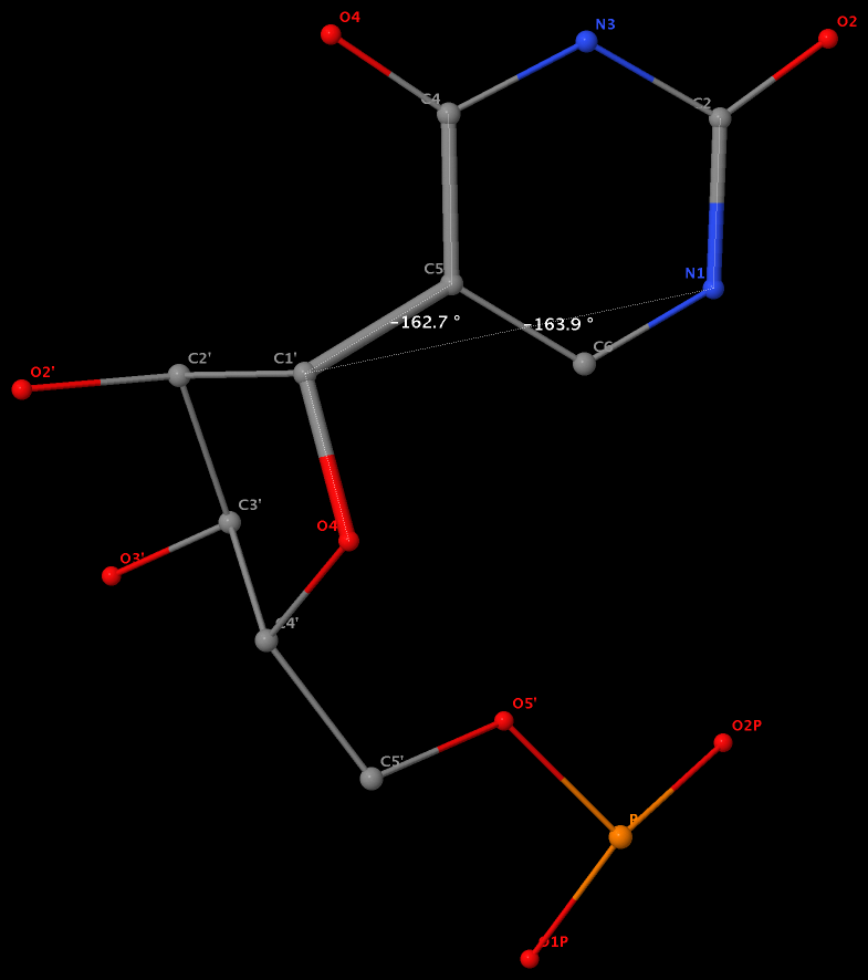
Pseudouridine (5-ribosyluracil, PSU) was the first identified modified nucleoside in RNA and is the most abundant. PSU is unique in that it has a C-glycosidic bond (C-C1′) instead of the N-glycosidic bond common to all other nucleosides, canonical or modified. It thus poses a problem as to how to calculate the χ torsion angle: should it be O4′-C1′-C5-C4, reflecting the actual glycosidic bond connection, or should the conventional definition O4′-C1′-N1-C2 still be applied literally? As a concrete example, the figure below shows the (slightly) different numerical values (–162.7° vs. –163.9°), as given by the two definitions, for PSU 6 on chain A of the PDB entry 3cgp (based on the 2009 Biochemistry article by Lin & Kielkopf titled X-ray structures of U2 snRNA-branchpoint duplexes containing conserved pseudouridines).
Needless to say, the specific definition of the χ torsion angle for PSU in RNA structures is a very subtle point, and I am not aware of any discussion on this issue in literature. In 3DNA, PSU is identified explicitly, and χ is defined by O4′-C1′-C5-C4. In NDB and a couple of other tools I am familiar with, χ for PSU is defined by O4′-C1′-N1-C2. Again using 3cgp (figure below) as an example, 3DNA gives –162.7°, whilst NDB gives –163.9°. Additionally, this distinction in N-C1′ vs. C-C1′ connection also comes into play when calculating the perpendicular distance from the 3′ phosphorus atom to the glycosidic bond, as per Richardson et al.
Except for pseudouridine, a nucleoside in DNA/RNA contains an N-glycosidic bond that connects the base to the sugar. The chi (χ) torsion angle, which characterizes the relative base/sugar orientation, is defined by O4′-C1′-N1-C2 for pyrimidines (C, T and U), and O4′-C1′-N9-C4 for purines (A and G).
Normally (as in A- and B-form DNA/RNA duplex), χ falls into the ranges of +90° to +180°; –90° to –180° (or 180° to 270°), corresponding to the anti conformation (Figure below, top). Occasionally, χ has values in the range of –90° to +90°, referring to the syn conformation (Figure below, bottom). Note that in left-handed Z-DNA with CG repeating sequence, the purine G is in syn conformation whilst the pyrimidine C is anti.
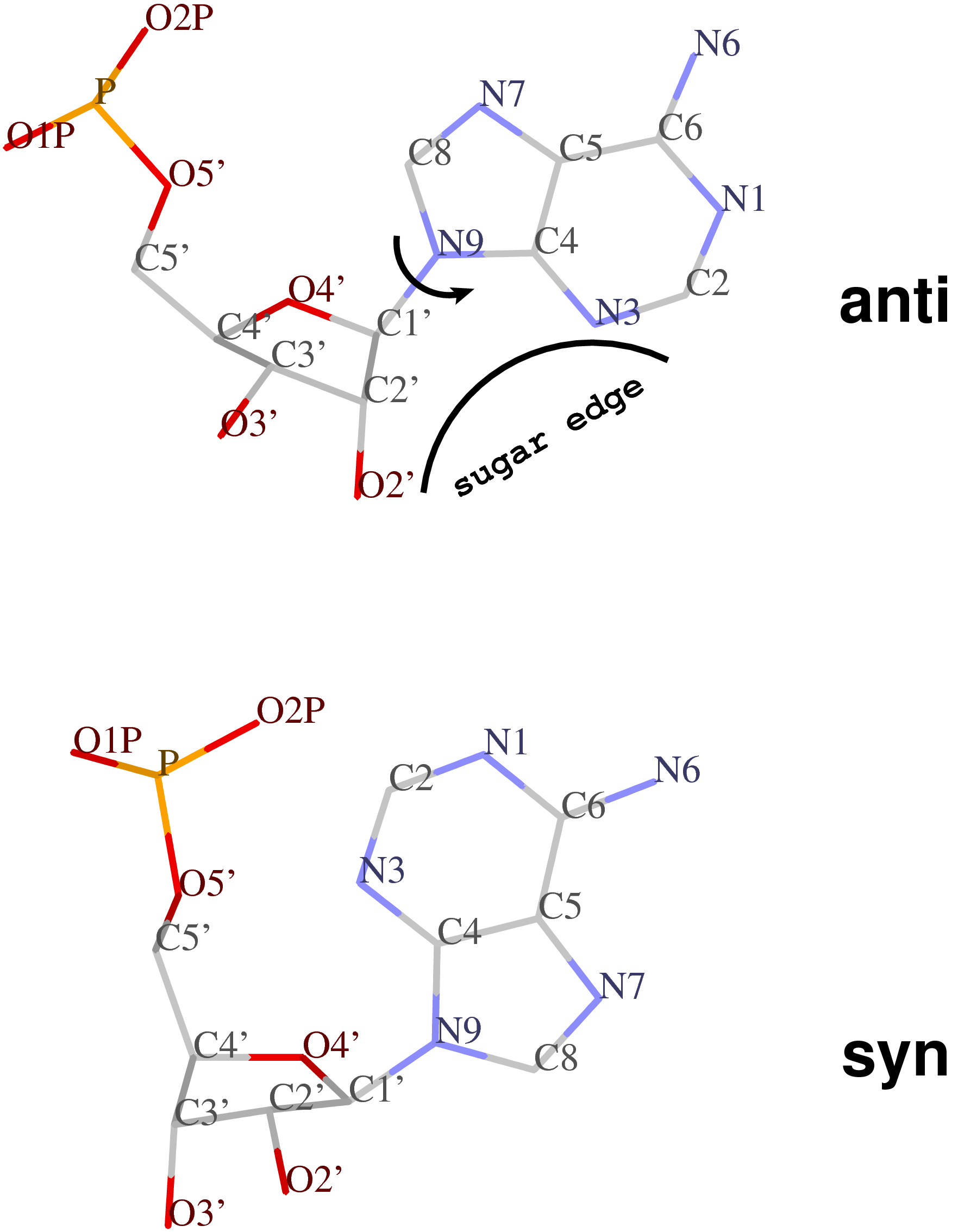
Presumably, the χ-related anti / syn conformation is a simple geometric concept. Nevertheless, the N-glycosidic bond and the corresponding χ torsion angle illustrate that the base and the sugar are two separate entities, i.e. there is an internal degree of freedom between them. In this respect, it is worth noting that the Leontis-Westhod sugar edge for base-pair classification corresponds to the anti form (as applied to RNA) only. When a base is flipped over into the syn conformation, the “sugar edge”, defined in connection with the minor (shallow) groove side of a nitrogenous bases, simply does not exist.
Base-flipping (anti / syn conformation switch) is one of the factors associated with the two possible relative orientations of the two bases in a pair, characterized explicitly in 3DNA as of type M+N or M–N since the 2003 NAR paper (Figure 2, linked below). I re-emphasized this distinction in our 2010 GpU dinucleotide platform paper (in particular, see supplementary Figure S2). Unfortunately, this subtle (but crucial, in my opinion) point has never been taken seriously (or at all) by the RNA community, even with 3DNA’s wide adoption. However, as people know 3DNA deeper/better and take RNA base-pair classification more rigorously, I have no doubt that the simplicity of this explicit distinction and the resultant full quantification of each and every possible base pair using standard geometric parameters will gradually be appreciated.

As of 3DNA v2.1, the output of the χ torsion angle is also associated with its classification in anti / syn conformation, among other new features (see for example the output for 6tna).