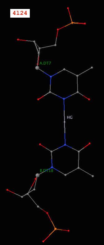
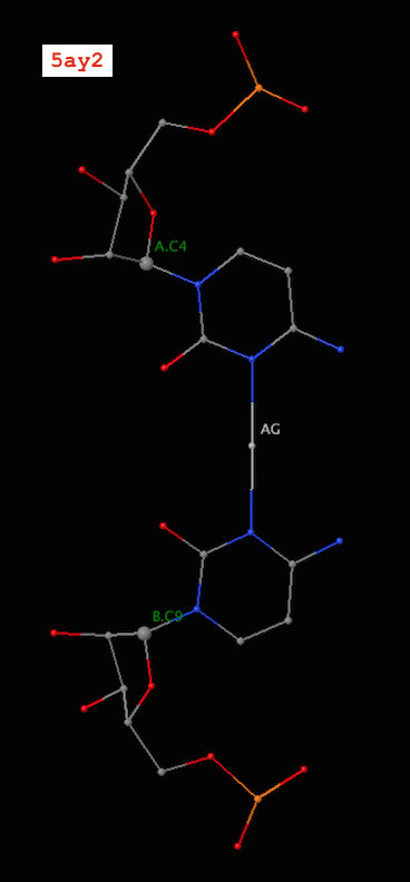
Recently, I became aware of the metallo-base pairs, such as T-Hg-T (PDB id: 4l24) and C-Ag-C (5ay2) from the work of Kondo et al (Pubmed: 24478025 and 26448329). As of v1.4.3-2015oct23, DSSR can detect such metallo-bps automatically, as shown below:
# x3dna-dssr -i=4l24.pdb -o=4l24.out
List of 12 base pairs
      nt1            nt2           bp  name        Saenger    LW  DSSR
   1 A.DC1          B.DG24         C-G WC          19-XIX    cWW  cW-W
   2 A.DG2          B.DC23         G-C WC          19-XIX    cWW  cW-W
   3 A.DC3          B.DG22         C-G WC          19-XIX    cWW  cW-W
   4 A.DG4          B.DC21         G-C WC          19-XIX    cWW  cW-W
   5 A.DA5          B.DT20         A-T WC          20-XX     cWW  cW-W
   6 A.DT6          B.DT19         T-T Metal       n/a       cWW  cW-W
   7 A.DT7          B.DT18         T-T Metal       n/a       cWW  cW-W
   8 A.DT8          B.DA17         T-A WC          20-XX     cWW  cW-W
   9 A.DC9          B.DG16         C-G WC          19-XIX    cWW  cW-W
  10 A.DG10         B.DC15         G-C WC          19-XIX    cWW  cW-W
  11 A.DC11         B.DG14         C-G WC          19-XIX    cWW  cW-W
  12 A.DG12         B.DC13         G-C WC          19-XIX    cWW  cW-W
and
# x3dna-dssr -i=5ay2.pdb -o=5ay2.out
List of 24 base pairs
      nt1            nt2           bp  name        Saenger    LW  DSSR
   1 A.G1           B.C12          G-C WC          19-XIX    cWW  cW-W
   2 A.G2           B.C11          G-C WC          19-XIX    cWW  cW-W
   3 A.A3           B.U10          A-U WC          20-XX     cWW  cW-W
   4 A.C4           B.C9           C-C Metal       n/a       cWW  cW-W
   5 A.U5           B.A8           U-A WC          20-XX     cWW  cW-W
   6 A.CBR6         B.G7           c-G WC          19-XIX    cWW  cW-W
   7 A.G7           B.CBR6         G-c WC          19-XIX    cWW  cW-W
   8 A.A8           B.U5           A-U WC          20-XX     cWW  cW-W
   9 A.C9           B.C4           C-C Metal       n/a       cWW  cW-W
  10 A.U10          B.A3           U-A WC          20-XX     cWW  cW-W
  11 A.C11          B.G2           C-G WC          19-XIX    cWW  cW-W
  12 A.C12          B.G1           C-G WC          19-XIX    cWW  cW-W
  13 C.G1           D.C12          G-C WC          19-XIX    cWW  cW-W
  14 C.G2           D.C11          G-C WC          19-XIX    cWW  cW-W
  15 C.A3           D.U10          A-U WC          20-XX     cWW  cW-W
  16 C.C4           D.C9           C-C Metal       n/a       cWW  cW-W
  17 C.U5           D.A8           U-A WC          20-XX     cWW  cW-W
  18 C.CBR6         D.G7           c-G WC          19-XIX    cWW  cW-W
  19 C.G7           D.CBR6         G-c WC          19-XIX    cWW  cW-W
  20 C.A8           D.U5           A-U WC          20-XX     cWW  cW-W
  21 C.C9           D.C4           C-C Metal       n/a       cWW  cW-W
  22 C.U10          D.A3           U-A WC          20-XX     cWW  cW-W
  23 C.C11          D.G2           C-G WC          19-XIX    cWW  cW-W
  24 C.C12          D.G1           C-G WC          19-XIX    cWW  cW-W
Note the name “Metal” for the metallo-bps. Moreover, the corresponding entries in the ‘dssr-pairs.pdb’ file also include the metal ions, as shown below:


It is worth noting that in a metallo-bp, the metal ion lies approximately in the bp plane. Moreover, it is in the middle of the two bases, which would otherwise not form a pair in the conventional sense.

Curves+ and 3DNA are currently the most widely used programs for analyzing nucleic acid structures (predominantly double helices). As noted in my blog post, Curves+ vs 3DNA, these two programs also complement each other in terms of features. It thus makes sense to run both to get a better understanding of the DNA/RNA structures one is interested in.
Indeed, over the past few years, I have seen quite a few articles citing both 3DNA and Curves+. Listed below are three recent examples:
The helical parameters were measured with 3DNA33 and Curves+.34 The local helical parameters are defined with regard to base steps and without regard to a global axis.
Structure analysis. Helix, base and base pair parameters were calculated with 3DNA or curve+ software packages23,24.
The major global difference between the native and mixed backbone structures is that the RNA backbone is compressed or kinked in strands containing the modified linkage (Fig. 3 B and C, by CURVES) (30). … To compare the three RNA structures at a more detailed and local level, we calculated the base pair helical and step parameters for all three structures using the 3DNA software tools (31) (Fig. 4 and Table S2). [In the Results section]
For each snapshot, the structural parameters—including six base pair parameters, six local base pair step parameters, and pseudorotation angles for each nucleotide—were calculated using 3DNA (31). The two terminal base pairs are omitted for the 3DNA analysis, because they unwind frequently in the triple 2′-5′-linked duplex. [In the Materials and Methods section]
Reading through these papers, however, it is not clear to me if the authors took advantage of the find_pair -curves+ option in 3DNA, as detailed in Building a bridge between Curves+ and 3DNA. Hopefully, this post will help draw more attention to this connection between Curves+ and 3DNA.

JSON (JavaScript Object Notation) is a simple human-readable format that expresses data objects in name-value pairs. Over the years, it has surpassed XML to become the preferred data exchange format between applications. As a result, I’ve recently added the --json command-line option to DSSR to make its numerous derived parameters easily accessible.
The DSSR JSON output is contained in a compact one-line text string that may look cryptic to the uninitiated. Yet, with commonly available JSON parsers or libraries, it is straightforward to make sense of the DSSR JSON output. In this blogpost, I am illustrating how to parse DSSR-derived .json file via two command-line tools, jq and Underscore-CLI.
jq — lightweight and flexible command-line JSON processor
According to its website,
jq is like sed for JSON data – you can use it to slice and filter and map and transform structured data with the same ease that sed, awk, grep and friends let you play with text.
Moreover, like DSSR per se, “jq is written in portable C, and it has zero runtime dependencies.” Prebuilt binaries are available for Linux, OS X and Windows. So it is trivial to get jq up and running. The current stable version is 1.5, released on August 15, 2015.
Using the crystal structure of yeast phenylalanine tRNA (1ehz) as an example, here are some sample usages with DSSR-derived JSON output:
    # Pretty print JSON
x3dna-dssr -i=1ehz.pdb --json | jq .
    # Extract the top-level keys, in insertion order
x3dna-dssr -i=1ehz.pdb --json | jq keys_unsorted
    # Extract parameters for nucleotides
x3dna-dssr -i=1ehz.pdb --json | jq .nts
    # Extract nucleotide id and its base reference frame
x3dna-dssr -i=1ehz.pdb --json | jq '.nts[] | (.nt_id, .frame)'
Underscore-CLI — command-line utility-belt for hacking JSON and Javascript.
Underscore-CLI is built upon Node.js, and can be installed using the npm package manager. It is claimed as ‘the “swiss-army-knife” tool for processing JSON data – can be used as a simple pretty-printer, or as a full-powered Javascript command-line.’
Following the above examples illustrating jq, here are the corresponding commands for Underscore-CLI:
x3dna-dssr -i=1ehz.pdb --json | underscore print --color
x3dna-dssr -i=1ehz.pdb --json | underscore keys --color
x3dna-dssr -i=1ehz.pdb --json | underscore select .nts --color
x3dna-dssr -i=1ehz.pdb --json | underscore select .nts | underscore select '.nt_id, .frame' --color
jq or Underscore-CLI — which one to use?
As always, it depends. While jq feels more like a standard Unix utility (as sed, awk, grep etc), Underscore-CLI is better integrated into the Javascript language. For simple applications such as parsing DSSR output, either jq or Underscore-CLI is more than sufficient.
I use jq most of the time, but resort to Underscore-CLI for its “smart whitespace”. Here is an example to illustrate the difference between the two:
# z-axis of A.G1 (1ehz) base reference frame
# jq output, split in 5 lines
    "z_axis": [
      0.799,
      0.488,
      -0.352
    ]
# Underscore-CLI, in a more-readable one line
    "z_axis": [0.799, 0.488, -0.352]

From early on, the x3dna.org domain and its related sub-domains (e.g., for the forum and the web-interface to DSSR) has been served via shared hosting. By and large, this simple arrangement has worked quite well. Over the years, though, I’ve gradually realized some of its inherent limitations. One is the limited resources available to the 3DNA-related websites. Another is the accessibility issue from countries like China.
To remedy such issues, I’ve recently moved the 3DNA Forum and the web-interface to DSSR to a dedicated web server at Columbia University. Moreover, a duplicate copy of the 3DNA homepage is made available via http://home.x3dna.org hosted at Columbia. The three new websites have been verified to be accessible directly from China.
These updates on x3dna.org not only ensure global accessibility to 3DNA/DSSR, but also allow for more web services to be made available.

As of v1.3.3-2015sep03, DSSR outputs the reference frame of any base or base-pair (bp). With an explicit list of such reference frames, one can better understand how the 3DNA/DSSR bp parameters are calculated. Moreover, third-party bioinformatics tools can take advantage of the frames for further exploration of nucleic acid structures, including visualization.
Let’s use the G1–C72 bp (detailed below) in the yeast phenylalanine tRNA (1ehz) as an example:
1 A.G1 A.C72 G-C WC 19-XIX cWW cW-W
The standard base reference frame for A.G1 is:
{
  rsmd: 0.008,
  origin: [53.757, 41.868, 52.93],
  x_axis: [-0.259, -0.25, -0.933],
  y_axis: [-0.543, 0.837, -0.073],
  z_axis: [0.799, 0.488, -0.352]
}
And the one for A.C72 is:
{
  rsmd: 0.006,
  origin: [53.779, 42.132, 52.224],
  x_axis: [-0.402, -0.311, -0.861],
  y_axis: [0.451, -0.886, 0.109],
  z_axis: [-0.797, -0.345, 0.497]
}
The G1–C72 bp reference frame is:
{
  rsmd: null,
  origin: [53.768, 42, 52.577],
  x_axis: [-0.331, -0.283, -0.9],
  y_axis: [-0.497, 0.863, -0.089],
  z_axis: [0.802, 0.418, -0.427]
}
The beauty of the DSSR JSON output is that the above information can be extracted on the fly. For example, the following commands extract the above frames:
x3dna-dssr -i=1ehz.pdb --json | jq '.ntParams[] | select(.nt_id=="A.G1") | .frame'
x3dna-dssr -i=1ehz.pdb --json | jq '.ntParams[] | select(.nt_id=="A.C72") | .frame'
x3dna-dssr -i=1ehz.pdb --json --more | jq .pairs[0].frame
Note that in JSON, the array is 0-indexed, so the first bp (G1–C72) has an index of 0. In addition to jq, I also used underscore to pretty-print the frames.

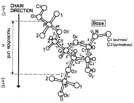
The conformation of the five-membered sugar ring in DNA/RNA structures can be characterized using the five corresponding endocyclic torsion angles (shown below).

v0: C4'-O4'-C1'-C2'
v1: O4'-C1'-C2'-C3'
v2: C1'-C2'-C3'-C4'
v3: C2'-C3'-C4'-O4'
v4: C3'-C4'-O4'-C1'
On account of the five-member ring constraint, the conformation can be characterized approximately by 5 - 3 = 2 parameters. Using the concept of pseudorotation of the sugar ring, the two parameters are the amplitude (τm) and phase angle (P, in the range of 0° to 360°).
One set of widely used formula to convert the five torsion angles to the pseudorotation parameters is due to Altona & Sundaralingam (1972): “Conformational Analysis of the Sugar Ring in Nucleosides and Nucleotides. A New Description Using the Concept of Pseudorotation” [J. Am. Chem. Soc., 94(23), pp 8205–8212]. As always, the concept is best illustrated with an example. Here I use the sugar ring of G4 (chain A) of the Dickerson-Drew dodecamer (1bna), with Matlab/Octave code:
# xyz coordinates of the sugar ring: G4 (chain A), 1bna
ATOM     63  C4'  DG A   4      21.393  16.960  18.505  1.00 53.00
ATOM     64  O4'  DG A   4      20.353  17.952  18.496  1.00 38.79
ATOM     65  C3'  DG A   4      21.264  16.229  17.176  1.00 56.72
ATOM     67  C2'  DG A   4      20.793  17.368  16.288  1.00 40.81
ATOM     68  C1'  DG A   4      19.716  17.901  17.218  1.00 30.52

# endocyclic torsion angles:
v0 = -26.7; v1 = 46.3; v2 = -47.1; v3 = 33.4; v4 = -4.4
Pconst = sin(pi/5) + sin(pi/2.5)  # 1.5388
P0 = atan2(v4 + v1 - v3 - v0, 2.0 * v2 * Pconst);  # 2.9034
tm = v2 / cos(P0);  # amplitude: 48.469
P = 180/pi * P0;  # phase angle: 166.35 [P + 360 if P0 < 0]
The Altona & Sundaralingam (1972) pseudorotation parameters are what have been adopted in 3DNA, following the NewHelix program of Dr. Dickerson. The Curves+ program, on the other hand, uses another (newer) set of formula due to Westhof & Sundaralingam (1983): “A Method for the Analysis of Puckering Disorder in Five-Membered Rings: The Relative Mobilities of Furanose and Proline Rings and Their Effects on Polynucleotide and Polypeptide Backbone Flexibility” [J. Am. Chem. Soc., 105(4), pp 970–976]. The two sets of formula, by Altona & Sundaralingam (1972) and Westhof & Sundaralingam (1983), give slightly different numerical values for the two pseudorotation parameters (τm and P).
Since 3DNA and Curves+ are currently two of the most widely used programs for conformational analysis of nucleic acid structures, the subtle differences in pseudorotation parameters may cause confusions for users who use (or are familiar with) both programs. Over the past few years, I have indeed received such questions via email.
With the same G4 (chain A, 1bna) sugar ring, here is the Matlab/Octave script showing how Curve+ calculates the pseudorotation parameters:
# xyz coordinates of sugar ring G4 (chain A, 1bna)

# endocyclic torsion angles, same as above
v0 = -26.7; v1 = 46.3; v2 = -47.1; v3 = 33.4; v4 = -4.4

v = [v2, v3, v4, v0, v1]; # reorder them into vector v[]
A = 0; B = 0;
for i = 1:5
    t = 0.8 * pi * (i - 1);
    A += v(i) * cos(t);
    B += v(i) * sin(t);
end
A *= 0.4;   # -48.476
B *= -0.4;  # 11.516

tm = sqrt(A * A + B * B);  # 49.825

c = A/tm; s = B/tm;
P = atan2(s, c) * 180 / pi;  # 166.64
For this specific example, i.e., the sugar ring of G4 (chain A, 1bna), the pseudorotation parameters as calculated by 3DNA per Altona & Sundaralingam (1972) and Curves+ per Westhof & Sundaralingam (1983) are as follows:
           amplitude        phase angle
3DNA        48.469             166.35
Curves+     49.825             166.64
Needless to say, the differences are subtle, and few people will notice/bother at all. For those who do care about such little details, however, hopefully this post will help you understand where the differences actually come from.
For consistency with the 3DNA output, DSSR (by default) also follows the Altona & Sundaralingam (1972) definitions of sugar pseudorotation. Nevertheless, DSSR also contains an undocumented option, --sugar-pucker=westhof83, to output τm and P according to the Westhof & Sundaralingam (1983) definitions.
Each sugar is assigned into one of the following ten puckering modes, by dividing the phase angle (P, in the range of 0° to 360°) into 36° ranges reach.
C3'-endo, C4'-exo,   O4'-endo, C1'-exo,  C2'-endo,
C3'-exo,  C4'-endo,  O4'-exo,  C1'-endo, C2'-exo
For sugars in nucleic acid structures, C3’-endo [0°, 36°) and C2’-endo [144°, 180°) are predominant. The former corresponds to sugars in ‘canonical’ RNA or A-form DNA, and the latter in sugars of standard B-form DNA. In reality, RNA structures as deposited in the PDB could also contain C2′-endo sugars. One significant example is the GpU dinucleotide platforms, where the 5′-ribose sugar (G) is in the C2′-endo form and the 3′-sugar (U) in the C3′-endo form — see my blog post, titled ‘Is the O2′(G)…O2P H-bond in GpU platforms real?’.
Notes:
- This post is based on my 2011-06-11 blog post with the same title.
- While visiting Lyon in July 2014, I had the opportunity to hear Dr. Lavery’s opinion on adopting the Westhof & Sundaralingam (1983) sugar-pucker definitions in Curves+. I learned that the new formula are more robust in rare, extreme cases of sugar conformation than the 1972 variants. After all, Dr. Sundaralingam is a co-author on both papers. It is possible that in future releases of DSSR, the new 1983 formula for sugar pucker would become the default.

In the DSSR v1.2.7-2015jun09 release, I documented two additional command-line options (--prefix and --cleanup) that are related to the various auxiliary files. As a matter of fact, these two options (among quite a few others) have been there for a long time, but without being explicitly described. The point is not to hide but to simplify — one of the design goals of DSSR is simplicity. DSSR has already possessed numerous key functionality to be appreciated. Before DSSR is firmly established in the RNA bioinformatics field, I beleive too many nonessential “features” could be distracting. While writing and refining the DSSR code, I do feel that some ‘auxiliary’ features could be handy for experienced users (including myself). So along the way, I’ve added many ‘hidden’ options that are either experimental or potentially useful.
On one side, I sense it is acceptable for a scientific software to actually does more than it claims. On the other hand, I have always been quick in addressing users’ requests — as one example, check for the --select option recently introduced into DSSR in response to a user request, and the ‘hidden’ --dbn-break option for specifying the symbol to separate multiple chains or chain breaks in DSSR-derived dot-bracket notation.
Back to --prefix and --cleanup, the purposes of these two closely related options can be best illustrated using the yeast phenylalanine tRNA structure (1ehz) as an example. By default, running x3dna-dssr -i=1ehz.pdb will produce a total of 11 auxiliary files, with names prefixed with dssr-, as shown below:
List of 11 additional files
   1 dssr-stems.pdb -- an ensemble of stems
   2 dssr-helices.pdb -- an ensemble of helices (coaxial stacking)
   3 dssr-pairs.pdb -- an ensemble of base pairs
   4 dssr-multiplets.pdb -- an ensemble of multiplets
   5 dssr-hairpins.pdb -- an ensemble of hairpin loops
   6 dssr-junctions.pdb -- an ensemble of junctions (multi-branch)
   7 dssr-2ndstrs.bpseq -- secondary structure in bpseq format
   8 dssr-2ndstrs.ct -- secondary structure in connect table format
   9 dssr-2ndstrs.dbn -- secondary structure in dot-bracket notation
  10 dssr-torsions.txt -- backbone torsion angles and suite names
  11 dssr-stacks.pdb -- an ensemble of stacks
With ‘fixed’ generic names by default, users can run DSSR in a directory repeatedly without creating too many files. This practice follows that used in the 3DNA suite of programs. However, my experience in supporting 3DNA over the years has shown that users (myself included) may want to explore further some of the files, e.g. ‘dssr-multiplets.pdb’ for displaying the base multiplets (four triplets here). One could easily use command-line (script) to change a generic name to a more appropriate one: e.g., mv dssr-multiplets.pdb 1ehz-multiplets.pdb for 1ehz. A better solution, however, is by introducing a customized prefix to the additional files, and that’s exactly where the --prefix option comes in. The option is specified like this: --prefix=text where text can be any string as appropriate. So running x3dna-dssr -i=1ehz.pdb --prefix=1ehz, for example, will lead to the following output:
List of 11 additional files
   1 1ehz-stems.pdb -- an ensemble of stems
   2 1ehz-helices.pdb -- an ensemble of helices (coaxial stacking)
   3 1ehz-pairs.pdb -- an ensemble of base pairs
   4 1ehz-multiplets.pdb -- an ensemble of multiplets
   5 1ehz-hairpins.pdb -- an ensemble of hairpin loops
   6 1ehz-junctions.pdb -- an ensemble of junctions (multi-branch)
   7 1ehz-2ndstrs.bpseq -- secondary structure in bpseq format
   8 1ehz-2ndstrs.ct -- secondary structure in connect table format
   9 1ehz-2ndstrs.dbn -- secondary structure in dot-bracket notation
  10 1ehz-torsions.txt -- backbone torsion angles and suite names
  11 1ehz-stacks.pdb -- an ensemble of stacks
The --cleanup option, as its name implies, is to tidy up a directory by removing the auxiliary files generated by DSSR. The usage is very simple:
x3dna-dssr --cleanup
x3dna-dssr --cleanup --prefix=1ehz
The former gets rid of the default ‘fixed’ generic auxiliary files (dssr-pairs.pdb etc), whilst the latter deletes prefixed supporting files (1ehz-pairs.pdb etc).

Recently, I came across and have been surprised by the different assignment of HETATM vs. ATOM records for modified nucleotides in PDB vs. PDBx/mmCIF format. As always, the issue is best illustrated with a concrete example. Here is what I observed in the PDB entry 1ehz, the crystal structure of yeast phenylalanine tRNA at 1.93 Å resolution.
DSSR identifies 14 modified nucleotides (of 11 types) in 1ehz as shown below:
List of 11 types of 14 modified nucleotides
      nt    count  list
   1 1MA-a    1    A.1MA58
   2 2MG-g    1    A.2MG10
   3 5MC-c    2    A.5MC40,A.5MC49
   4 5MU-t    1    A.5MU54
   5 7MG-g    1    A.7MG46
   6 H2U-u    2    A.H2U16,A.H2U17
   7 M2G-g    1    A.M2G26
   8 OMC-c    1    A.OMC32
   9 OMG-g    1    A.OMG34
  10 PSU-P    2    A.PSU39,A.PSU55
  11 YYG-g    1    A.YYG37
In file 1ehz.pdb downloaded from RCSB PDB, all the 14 modified nucleotides are assigned as HETATM whereas in 1ehz.cif the corresponding records are ATOM. Here is the excerpt for 1MA58 in PDB format:
HETATM 1252  P   1MA A  58      73.770  67.765  34.057  1.00 30.65           P
HETATM 1253  OP1 1MA A  58      72.638  67.886  33.105  1.00 32.84           O
HETATM 1254  OP2 1MA A  58      73.621  68.229  35.450  1.00 29.49           O
HETATM 1255  O5' 1MA A  58      74.315  66.273  34.254  1.00 28.81           O
HETATM 1256  C5' 1MA A  58      74.592  65.439  33.080  1.00 29.42           C
HETATM 1257  C4' 1MA A  58      74.279  63.972  33.383  1.00 33.42           C
HETATM 1258  O4' 1MA A  58      74.880  63.685  34.667  1.00 32.36           O
HETATM 1259  C3' 1MA A  58      72.789  63.573  33.509  1.00 35.13           C
HETATM 1260  O3' 1MA A  58      72.625  62.168  33.250  1.00 36.80           O
HETATM 1261  C2' 1MA A  58      72.560  63.667  35.012  1.00 34.80           C
HETATM 1262  O2' 1MA A  58      71.525  62.828  35.506  1.00 36.27           O
HETATM 1263  C1' 1MA A  58      73.908  63.150  35.551  1.00 33.62           C
HETATM 1264  N9  1MA A  58      74.284  63.494  36.930  1.00 30.36           N
HETATM 1265  C8  1MA A  58      73.887  64.574  37.688  1.00 34.55           C
HETATM 1266  N7  1MA A  58      74.415  64.610  38.899  1.00 33.32           N
HETATM 1267  C5  1MA A  58      75.204  63.469  38.953  1.00 33.37           C
HETATM 1268  C6  1MA A  58      76.031  62.941  39.948  1.00 33.58           C
HETATM 1269  N6  1MA A  58      76.184  63.488  41.134  1.00 41.19           N
HETATM 1270  N1  1MA A  58      76.708  61.803  39.669  1.00 34.48           N
HETATM 1271  CM1 1MA A  58      77.649  61.222  40.626  1.00 31.43           C
HETATM 1272  C2  1MA A  58      76.527  61.216  38.479  1.00 28.43           C
HETATM 1273  N3  1MA A  58      75.793  61.624  37.453  1.00 31.67           N
HETATM 1274  C4  1MA A  58      75.142  62.771  37.747  1.00 33.02           C
The corresponding section in PDBx/mmCIF format is:
ATOM   1252 P  P     . 1MA A 1 58 ? 73.770 67.765 34.057  1.00 30.65  ? ? ? ? ? ? 58  1MA A P     1
ATOM   1253 O  OP1   . 1MA A 1 58 ? 72.638 67.886 33.105  1.00 32.84  ? ? ? ? ? ? 58  1MA A OP1   1
ATOM   1254 O  OP2   . 1MA A 1 58 ? 73.621 68.229 35.450  1.00 29.49  ? ? ? ? ? ? 58  1MA A OP2   1
ATOM   1255 O  "O5'" . 1MA A 1 58 ? 74.315 66.273 34.254  1.00 28.81  ? ? ? ? ? ? 58  1MA A "O5'" 1
ATOM   1256 C  "C5'" . 1MA A 1 58 ? 74.592 65.439 33.080  1.00 29.42  ? ? ? ? ? ? 58  1MA A "C5'" 1
ATOM   1257 C  "C4'" . 1MA A 1 58 ? 74.279 63.972 33.383  1.00 33.42  ? ? ? ? ? ? 58  1MA A "C4'" 1
ATOM   1258 O  "O4'" . 1MA A 1 58 ? 74.880 63.685 34.667  1.00 32.36  ? ? ? ? ? ? 58  1MA A "O4'" 1
ATOM   1259 C  "C3'" . 1MA A 1 58 ? 72.789 63.573 33.509  1.00 35.13  ? ? ? ? ? ? 58  1MA A "C3'" 1
ATOM   1260 O  "O3'" . 1MA A 1 58 ? 72.625 62.168 33.250  1.00 36.80  ? ? ? ? ? ? 58  1MA A "O3'" 1
ATOM   1261 C  "C2'" . 1MA A 1 58 ? 72.560 63.667 35.012  1.00 34.80  ? ? ? ? ? ? 58  1MA A "C2'" 1
ATOM   1262 O  "O2'" . 1MA A 1 58 ? 71.525 62.828 35.506  1.00 36.27  ? ? ? ? ? ? 58  1MA A "O2'" 1
ATOM   1263 C  "C1'" . 1MA A 1 58 ? 73.908 63.150 35.551  1.00 33.62  ? ? ? ? ? ? 58  1MA A "C1'" 1
ATOM   1264 N  N9    . 1MA A 1 58 ? 74.284 63.494 36.930  1.00 30.36  ? ? ? ? ? ? 58  1MA A N9    1
ATOM   1265 C  C8    . 1MA A 1 58 ? 73.887 64.574 37.688  1.00 34.55  ? ? ? ? ? ? 58  1MA A C8    1
ATOM   1266 N  N7    . 1MA A 1 58 ? 74.415 64.610 38.899  1.00 33.32  ? ? ? ? ? ? 58  1MA A N7    1
ATOM   1267 C  C5    . 1MA A 1 58 ? 75.204 63.469 38.953  1.00 33.37  ? ? ? ? ? ? 58  1MA A C5    1
ATOM   1268 C  C6    . 1MA A 1 58 ? 76.031 62.941 39.948  1.00 33.58  ? ? ? ? ? ? 58  1MA A C6    1
ATOM   1269 N  N6    . 1MA A 1 58 ? 76.184 63.488 41.134  1.00 41.19  ? ? ? ? ? ? 58  1MA A N6    1
ATOM   1270 N  N1    . 1MA A 1 58 ? 76.708 61.803 39.669  1.00 34.48  ? ? ? ? ? ? 58  1MA A N1    1
ATOM   1271 C  CM1   . 1MA A 1 58 ? 77.649 61.222 40.626  1.00 31.43  ? ? ? ? ? ? 58  1MA A CM1   1
ATOM   1272 C  C2    . 1MA A 1 58 ? 76.527 61.216 38.479  1.00 28.43  ? ? ? ? ? ? 58  1MA A C2    1
ATOM   1273 N  N3    . 1MA A 1 58 ? 75.793 61.624 37.453  1.00 31.67  ? ? ? ? ? ? 58  1MA A N3    1
ATOM   1274 C  C4    . 1MA A 1 58 ? 75.142 62.771 37.747  1.00 33.02  ? ? ? ? ? ? 58  1MA A C4    1
While I have not tested exhaustively, it seems true that PDBx/mmCIF has adopted a different definition of what constitutes a HETATM residue. It is worth noting that results from 3DNA and DSSR/SNAP are not effected by the conflicting assignments.

From the very beginning, 3DNA contains two key programs, analyze and rebuild, for the analysis and rebuilding of nucleic acid 3D structures. The two names are short and to the point, but with one caveat. They are common verbs that can be easily picked up by other software packages. When 3DNA and such packages are installed on the same machine, naming clashes happen. If the 3DNA bin/ directory is searched afterwards, the analyze or rebuild command may have nothing to do with nucleic acid structures at all. Naturally, this naming ambiguity can lead to confusions and frustrations.
I’ve been aware of the rebuild program name conflict for a long time. Recently, I was surprised by another analyze program on my Mac OS X Yosemite. As shown from the following output, the analyze program seems to be installed via Mac port, and it is about analyzing words in a dictionary file.
~ [540] which analyze
/opt/local/bin/analyze
~ [541] analyze -h
correct syntax is:
analyze affix_file dictionary_file file_of_words_to_check
use two words per line for morphological generation
The ambiguous names are exactly the reason that I use x3dna-dssr and x3dna-snap for the two new programs I’ve been working over the past few years. As for the analyze and rebuild programs in 3DNA v2.x, I’d rather leave them as is. 3DNA is now in wide use in other structural bioinformatic pipelines to allow for easy name changes without causing compatibility issues. On a positive side, once you know the problem, fixing it is straightforward. This post is to raise the awareness of the 3DNA user community about such naming conflicts.


