An abasic site is a location in DNA or RNA where a purine or pyrimidine base is missing. It is also termed an AP site (i.e., apurinic/apyrimidinic site) in biochemistry and molecular genetics. The abasic site can be formed either spontaneously (e.g., depurination) or due to DNA damage (occurring as intermediates in base excision repair). According to Wikipedia, “It has been estimated that under physiological conditions 10,000 apurinic sites and 500 apyrimidinic may be generated in a cell daily.”
In DSSR and 3DNA v2.x, nucleotides are recognized using standard atom names and base planarity. Thus, abasic sites are not taken as nucleotides (by default), simply because they do not have base atoms. DSSR introduced the --abasic option to account for abasic sites, a feature useful for detecting loops with backbone connectivity.
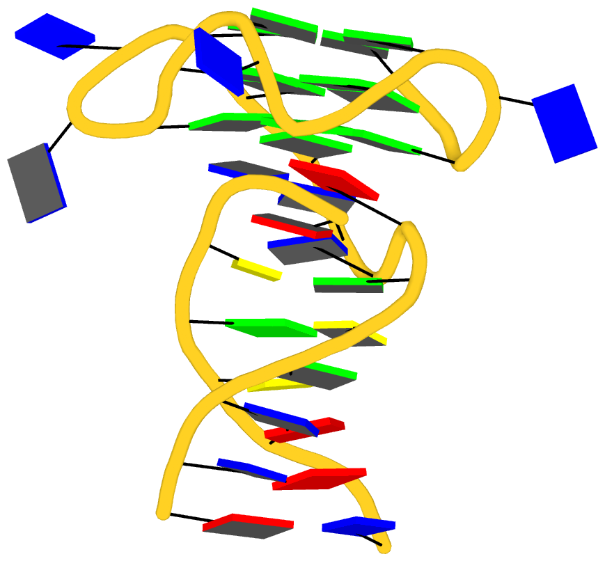
For example, by default, DSSR identifies one internal loop (no. 1 in the list below) in PDB entry 1l2c. With the --abasic option, two internal loops (including the one with the abasic site C.HPD18, no. 2) are detected.
List of 2 internal loops
   1 symmetric internal loop: nts=6; [1,1]; linked by [#-1,#1]
     summary: [2] 1 1 [B.1 C.24 B.3 C.22] 1 4
     nts=6 GTATAC B.DG1,B.DT2,B.DA3,C.DT22,C.DA23,C.DC24
       nts=1 T B.DT2
       nts=1 A C.DA23
   2 symmetric internal loop: nts=6; [1,1]; linked by [#1,#2]
     summary: [2] 1 1 [B.6 C.19 B.8 C.17] 4 5
     nts=6 CTTA?G B.DC6,B.DT7,B.DT8,C.DA17,C.HPD18,C.DG19
       nts=1 T B.DT7
       nts=1 ? C.HPD18
Note that C.HPD18 in 1l2c is a non-standard residue, as shown in the HETATM records below. Since the identity of C.HPD18 cannot be deduced from the atomic records, its one-letter code is designated as ?.
HETATM  346  P   HPD C  18     -14.637  52.299  29.949  1.00 49.12           P
HETATM  347  O5' HPD C  18     -14.658  52.173  28.359  1.00 48.28           O
HETATM  348  O1P HPD C  18     -15.167  51.040  30.537  1.00 49.35           O
HETATM  349  O2P HPD C  18     -13.303  52.798  30.369  1.00 46.43           O
HETATM  350  C5' HPD C  18     -15.703  51.469  27.687  1.00 45.70           C
HETATM  351  O4' HPD C  18     -16.364  50.501  25.561  1.00 44.15           O
HETATM  352  O3' HPD C  18     -13.990  51.738  24.335  1.00 45.75           O
HETATM  353  C1' HPD C  18     -16.105  54.187  25.684  1.00 52.47           C
HETATM  354  O1' HPD C  18     -17.309  54.085  26.496  1.00 56.16           O
HETATM  355  C3' HPD C  18     -14.756  52.250  25.426  1.00 46.23           C
HETATM  356  C4' HPD C  18     -15.263  51.093  26.291  1.00 45.72           C
HETATM  357  C2' HPD C  18     -16.030  52.889  24.898  1.00 49.05           C
In contrast, the R.U-8 in PDB entry 4ifd is a standard U, and is properly labeled by DSSR.
ATOM  26418  P     U R  -8     139.362  21.962 129.430  1.00208.29           P
ATOM  26419  OP1   U R  -8     140.062  20.821 130.074  1.00207.30           O
ATOM  26420  OP2   U R  -8     140.113  23.208 129.129  1.00208.44           O1+
ATOM  26421  O5'   U R  -8     138.712  21.439 128.071  1.00157.60           O
ATOM  26422  C5'   U R  -8     139.507  20.790 127.087  1.00155.47           C
ATOM  26423  C4'   U R  -8     138.843  20.804 125.731  1.00152.27           C
ATOM  26424  O4'   U R  -8     138.538  22.172 125.352  1.00149.29           O
ATOM  26425  C3'   U R  -8     139.677  20.275 124.572  1.00152.70           C
ATOM  26426  O3'   U R  -8     139.670  18.859 124.478  1.00155.04           O
ATOM  26427  C2'   U R  -8     139.053  20.969 123.369  1.00150.26           C
ATOM  26428  O2'   U R  -8     137.849  20.322 122.984  1.00146.83           O
ATOM  26429  C1'   U R  -8     138.700  22.334 123.958  1.00147.35           C
This is yet another little detail that DSSR takes care of. It is the close consideration to many such subtle points that makes DSSR different. Overall, DSSR represents my view of what a scientific software program could be (or should be).

Recently, while analyzing a representative set of RNA structures from the PDB, I came across three weird entries. They are documented below, primarily for my own record.
- 5els — “Structure of the KH domain of T-STAR in complex with AAAUAA RNA”. There are two alternative conformations for the six-nt
AAAUAA RNA component, labeled A and B, respectively. Normally, the A/B alternative coordinates for each atom are put directly next to each other, and assigned the same chain id, as in 1msy for the phosphate group of G2669 on chain A. In 5els, however, the two alternative conformations (A/B) are separated into two chains: chain H for A, and chain I for B.
- 1vql — “The structure of the transition state analogue ‘DCSN’ bound to the large ribosomal subunit of Haloarcula marismortui”. The three-nt fragment DA179—C180—C181 on chain 4 is in the 3’—>5’ direction.
- 4r3i — “The crystal structure of m(6)A RNA with the YTHDC1 YTH domain”. The mmCIF file has a model number of 0, instead of 1 (as in other cases I am aware of).

3DNA contains 55 fiber models compiled from literature, plus a derived RNA model (as of v2.1). To the best of my knowledge, this is the most comprehensive collection of regular DNA/RNA models. Please see Table 4 of the 2003 3DNA NAR paper for detailed structural features of these models and references.
The 55 models are based on the following works:
- Chandrasekaran & Arnott (from #1 to #43) — the most well-known set of fiber models
- Alexeev et al. (#44-#45)
- van Dam & Levitt (#46-#47)
- Premilat & Albiser (#48-#55)
The utility program fiber makes the generation of all these fiber models in a simple, consistent interface, and produces coordinate files in either PDB or PDBML format. Of those models, some can be built with an arbitrary sequence of A, C, G and T (e.g., A-/B-/C-DNA from calf thymus), while others are of fixed sequences (e.g., Z-DNA with GC repeats). The sequence can be specified either from command-line or a plain text file, in either lower, UPPER, or MixED cases.
Once 3DNA in properly installed, the command-line interface is the most versatile and convenient way to generate, e.g., a regular double-stranded DNA (mostly, B-DNA) of arbitrary sequence. The command-help message (generated with fiber -h) is as below:
NAME
        fiber - generate 55 fiber models based on Arnott and other's work
SYNOPSIS
        fiber [OPTION] PDBFILE
DESCRIPTION
        generate 55 fiber models based on the repeating unit from Arnott's
        work, including the canonical A-, B-, C- and Z-DNA, triplex, etc
        -xml     output structure coordinates in PDBML format
        -num     a structure identification number in the range (1-55)
        -m, -l   brief description of the 55 fiber structures
        -a, -1   A-DNA model (calf thymus)
        -b, -4   B-DNA (calf thymus, default)
        -c, -47  C-DNA (BII-type nucleotides)
        -d, -48  D(A)-DNA  ploy d(AT) : ploy d(AT) (right-handed)
        -z, -15  Z-DNA poly d(GC) : poly d(GC)
        -rna     for RNA with arbitrary base sequence
        -seq=string specifying an arbitrary base sequence
        -single  output a single-stranded structure
        -h       this help message (any non-recognized options will do)
INPUT
        An structural identification number (symbol)
EXAMPLES
        fiber fiber-BDNA.pdb
            # fiber -4 fiber-BDNA.pdb
            # fiber -b fiber-BDNA.pdb
        fiber -a fiber-ADNA.pdb
        fiber -seq=AAAGGUUU -rna fiber-RNA.pdb
        fiber -seq=AAAGGUUU -rna -single fiber-ssRNA.pdb
OUTPUT
        PDB file
SEE ALSO
        analyze, anyhelix, find_pair
AUTHOR
        3DNA v2.3-2016sept06, created and maintained by Xiang-Jun Lu (PhD)

Please post questions/comments on the 3DNA Forum: http://forum.x3dna.org/
Moreover, the w3DNA, 3D-DART web-interfaces, and the PyMOL wrapper make it easy to generate a regular DNA (or RNA) model, especially for occasional users or for educational purposes.
In principle, nothing is worth showing off with regard to 3DNA’s fiber model generation functionality. Nevertheless, this handy tool serves as a clear example of the differences between a “proof of concept” and a pragmatic software application. I initially decided to work on this tool simply for my own convenience. At that time, I had access to A-DNA and B-DNA fiber model generators, each as a separate program. Moreover, the constructed models did not comply to the PDB format in atom naming, among other subtitles.
I started with the Chandrasekaran & Arnott fiber models which I had a copy of data files. However, there were many details to work out, typos to correct, etc. to put them in a consistent framework. For other models, I had to read each original publication, and to type raw atomic cylindrical coordinates into computer. Again, quite a few inconsistencies popped up between the different publications with a time span over decades.
Overall, it was a quite tedious undertaking, requiring great attention to details. I am glad that I did that: I learned so much from the process, and more importantly, others can benefit from my effort. As I put in the 3DNA Nature Protocol paper (BOX 6 | FIBER-DIFFRACTION MODELS),
In preparing this set of fiber models, we have taken great care to ensure the accuracy and consistency of the models. For completeness and user verification, 3DNA includes, in addition to 3DNA-processed files, the original coordinates collected from the literature.
For those who want to understand what’s going on under the hood, there is no better way than to try to reproduce the process using, e.g., fiber B-DNA as an example.
From the very beginning, I had expected the 3DNA fiber functionality to serve as a handy tool for building a regular DNA duplex of chosen sequence. Over the years, the fiber program has gradually attracted attention from the community. The recent PyMOL wrapper by Thomas Holder is a clear sign of its increased popularity, and has prompted me to write this post, adapted largely from the one titled Fiber models in 3DNA make it easy to build regular DNA helices (dated Friday, October 9, 2009).
See also PyMOL wrapper to 3DNA fiber models

Given below is the content of the README file for fiber models in 3DNA:
1. The repeating units of each fiber structure are mostly based on the
   work of Chandrasekaran & Arnott (from #1 to #43). More recent fiber
   models are based on Alexeev et al. (#44-#45), van Dam & Levitt (#46
   -#47) and Premilat & Albiser (#48-#55).

2. Clean up of each residue
   a. currently ignore hydrogen atoms [can be easily added]
   b. change ME/C7 group of thymine to C5M
   c. re-assign O3' atom to be attached with C3'
   d. change distance unit from nm to A [most of the entries]
   e. re-ordering atoms according to the NDB convention

3. Fix up of problem structures.
   a. str#8 has no N9 atom for guanine
   b. str#10 is not available from the disk, manually input
   c. str#14 C5M atom was named C5 for Thymine, resulting two C5 atoms
   d. str#17 has wrong assignment of O3' atom on Guanine
   e. str#33 has wrong C6 position in U3
   f. str#37 to #str41 were typed in manually following Arnott's
        new list as given in "Oxford Handbook of Nucleic Acid Structure"
        edited by S. Neidle (Oxford Press, 1999)
   g. str#38 coordinates for N6(A) and N3(T) are WRONG as given in the
        original literature
   h. str#39 and #40 have the same O3' coordinates for the 2nd strand

4. str#44 & 45 have fixed strand II residues (T)

5. str#46 & 47 have +z-axis upwards (based on BI.pdb & BII.pdb)

6. str#48 to 55 have +z-axis upwards

List of 55 fiber structures

id#  Twist   Rise        Structure description
    (dgrees)  (A)
-------------------------------------------------------------------------------
 1   32.7   2.548  A-DNA  (calf thymus; generic sequence: A, C, G and T)
 2   65.5   5.095  A-DNA  poly d(ABr5U) : poly d(ABr5U)
 3    0.0  28.030  A-DNA  (calf thymus) poly d(A1T2C3G4G5A6A7T8G9G10T11) :
                                        poly d(A1C2C3A4T5T6C7C8G9A10T11)
 4   36.0   3.375  B-DNA  (calf thymus; generic sequence: A, C, G and T)
 5   72.0   6.720  B-DNA  poly d(CG) : poly d(CG)
 6  180.0  16.864  B-DNA  (calf thymus) poly d(C1C2C3C4C5) : poly d(G6G7G8G9G10)
 7   38.6   3.310  C-DNA  (calf thymus; generic sequence: A, C, G and T)
 8   40.0   3.312  C-DNA  poly d(GGT) : poly d(ACC)
 9  120.0   9.937  C-DNA  poly d(G1G2T3) : poly d(A4C5C6)
10   80.0   6.467  C-DNA  poly d(AG) : poly d(CT)
11   80.0   6.467  C-DNA  poly d(A1G2) : poly d(C3T4)
12   45.0   3.013  D-DNA  poly d(AAT) : poly d(ATT)
13   90.0   6.125  D-DNA  poly d(CI) : poly d(CI)
14  -90.0  18.500  D-DNA  poly d(A1T2A3T4A5T6) : poly d(A1T2A3T4A5T6)
15  -60.0   7.250  Z-DNA  poly d(GC) : poly d(GC)
16  -51.4   7.571  Z-DNA  poly d(As4T) : poly d(As4T)
17    0.0  10.200  L-DNA  (calf thymus) poly d(GC) : poly d(GC)
18   36.0   3.230  B'-DNA alpha poly d(A) : poly d(T) (H-DNA)
19   36.0   3.233  B'-DNA beta2 poly d(A) : poly d(T) (H-DNA  beta)
20   32.7   2.812  A-RNA  poly (A) : poly (U)
21   30.0   3.000  A'-RNA poly (I) : poly (C)
22   32.7   2.560  Hybrid poly (A) : poly d(T)
23   32.0   2.780  Hybrid poly d(G) : poly (C)
24   36.0   3.130  Hybrid poly d(I) : poly (C)
25   32.7   3.060  Hybrid poly d(A) : poly (U)
26   36.0   3.010  10-fold poly (X) : poly (X)
27   32.7   2.518  11-fold poly (X) : poly (X)
28   32.7   2.596  Poly (s2U) : poly (s2U) (symmetric base-pair)
29   32.7   2.596  Poly (s2U) : poly (s2U) (asymmetric base-pair)
30   32.7   3.160  Poly d(C) : poly d(I) : poly d(C)
31   30.0   3.260  Poly d(T) : poly d(A) : poly d(T)
32   32.7   3.040  Poly (U) : poly (A) : poly(U) (11-fold)
33   30.0   3.040  Poly (U) : poly (A) : poly(U) (12-fold)
34   30.0   3.290  Poly (I) : poly (A) : poly(I)
35   31.3   3.410  Poly (I) : poly (I) : poly(I) : poly(I)
36   60.0   3.155  Poly (C) or poly (mC) or poly (eC)
37   36.0   3.200  B'-DNA beta2  Poly d(A) : poly d(U)
38   36.0   3.240  B'-DNA beta1  Poly d(A) : poly d(T)
39   72.0   6.480  B'-DNA beta2  Poly d(AI) : poly d(CT)
40   72.0   6.460  B'-DNA beta1  Poly d(AI) : poly d(CT)
41  144.0  13.540  B'-DNA  Poly d(AATT) : poly d(AATT)
42   32.7   3.040  Poly(U) : poly d(A) : poly(U) [cf. #32]
43   36.0   3.200  Beta Poly d(A) : Poly d(U) [cf. #37]
44   36.0   3.233  Poly d(A) : poly d(T) (Ca salt)
45   36.0   3.233  Poly d(A) : poly d(T) (Na salt)
46   36.0   3.38   B-DNA (BI-type nucleotides; generic sequence: A, C, G and T)
47   40.0   3.32   C-DNA (BII-type nucleotides; generic sequence: A, C, G and T)
48   87.8   6.02   D(A)-DNA  ploy d(AT) : ploy d(AT) (right-handed)
49   60.0   7.20   S-DNA  ploy d(CG) : poly d(CG) (C_BG_A, right-handed)
50   60.0   7.20   S-DNA  ploy d(GC) : poly d(GC) (C_AG_B, right-handed)
51   31.6   3.22   B*-DNA  poly d(A) : poly d(T)
52   90.0   6.06   D(B)-DNA  poly d(AT) : poly d(AT) [cf. #48]
53  -38.7   3.29   C-DNA (generic sequence: A, C, G and T) (depreciated)
54   32.73  2.56   A-DNA (generic sequence: A, C, G and T) [cf. #1]
55   36.0   3.39   B-DNA (generic sequence: A, C, G and T) [cf. #4]
-------------------------------------------------------------------------------
List 1-41 based on Struther Arnott: ``Polynucleotide secondary structures:
     an historical perspective'', pp. 1-38 in ``Oxford Handbook of Nucleic
     Acid Structure'' edited by Stephen Neidle (Oxford Press, 1999).

     #42 and #43 are from Chandrasekaran & Arnott: "The Structures of DNA
     and RNA Helices in Oriented Fibers", pp 31-170 in "Landolt-Bornstein
     Numerical Data and Functional Relationships in Science and Technology"
     edited by W. Saenger (Springer-Verlag, 1990).

#44-#45 based on Alexeev et al., ``The structure of poly(dA) . poly(dT)
     as revealed by an X-ray fiber diffraction''. J. Biomol. Str. Dyn, 4,
     pp. 989-1011, 1987.

#46-#47 based on van Dam & Levitt, ``BII nucleotides in the B and C forms
     of natural-sequence polymeric DNA: a new model for the C form of DNA''.
     J. Mol. Biol., 304, pp. 541-561, 2000.

#48-#55 based on Premilat & Albiser, ``A new D-DNA form of poly(dA-dT) .
     poly(dA-dT): an A-DNA type structure with reversed Hoogsteen Pairing''.
     Eur. Biophys. J., 30, pp. 404-410, 2001 (and several other publications).

Recently, I heard from Thomas Holder, the PyMOL Principal Developer (Schrödinger, Inc.), that he had written a wrapper to the 3DNA fiber command. This PyMOL wrapper is implemented as part of his versatile PSICO library (see the PyMOL Wiki page Psico for details), and exposes the 55 fiber models based on Arnott and other’s work to the wide PyMOL user community. Moreover, the wrapper can be accessed directly from PyMOL (without installing PSICO), as shown below with an example:
PyMOL> run https://raw.githubusercontent.com/speleo3/pymol-psico/master/psico/creating.py
PyMOL> fiber CTAGCG
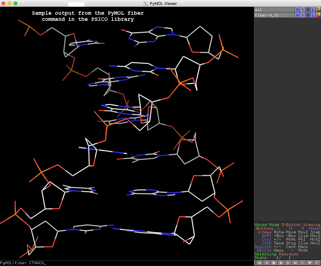
The resulting fiber model is the default B-form DNA of calf thymus, with twist of 36.0° and rise of 3.375 Å (see figure below). Note that cases in base sequence do not matter, so fiber ctagcg or fiber CTAgcg will give the same result.
Running PyMOL>help fiber gives the following detailed usages info, which should be sufficient to get one started with this fiber tool in PyMOL.
PyMOL> help fiber

DESCRIPTION

    Run X3DNA's "fiber" tool.

    For the list of structure identification numbers, see for example:
    http://xiang-jun.blogspot.com/2009/10/fiber-models-in-3dna.html

USAGE

    fiber seq [, num [, name [, rna [, single ]]]]

ARGUMENTS

    seq = str: single letter code sequence or number of repeats for
    repeat models.

    num = int: structure identification number {default: 4}

    name = str: name of object to create {default: random unused name}

    rna = 0/1: 0=DNA, 1=RNA {default: 0}

    single = 0/1: 0=double stranded, 1=single stranded {default: 0}

EXAMPLES

    # environment (this could go into ~/.pymolrc or ~/.bashrc)
    os.environ["X3DNA"] = "/opt/x3dna-v2.3"

    # B or A DNA from sequence
    fiber CTAGCG
    fiber CTAGCG, 1, ADNA

    # double or single stranded RNA from sequence
    fiber AAAGGU, name=dsRNA, rna=1
    fiber AAAGGU, name=ssRNA, rna=1, single=1

    # poly-GC Z-DNA repeat model with 10 repeats
    fiber 10, 15
Thanks to Thomas, for making another connection between PyMOL and 3DNA/DSSR. The other one is the DSSR-plugin for PyMOL to create “block” shaped cartoons for nucleic acid bases and base pairs.
See also 3DNA fiber models

Recently I read the article titled Structural Insights into the Quadruplex−Duplex 3′ Interface Formed from a Telomeric Repeat: A Potential Molecular Target by Krauss et al.. I quickly ran DSSR on the corresponding PDB entry is 5dww. Not surprisingly, DSSR can automatically identify reported key structural features (see output file 5dww.out for details), including the TAT triplet at the quadruplex−duplex junction, and the three G-quartets. Note that the result is based on biological assembly 1 in PDB file 5dww.pdb1 since the asymmetric unit contains four such molecules.
List of 4 multiplets
   1 nts=3 TAT 1:A.DT17,1:A.DA19,1:B.DT7
   2 nts=4 GGGG 1:A.DG1,1:A.DG5,1:A.DG9,1:A.DG14
   3 nts=4 GGGG 1:A.DG2,1:A.DG6,1:A.DG10,1:A.DG15
   4 nts=4 GGGG 1:A.DG3,1:A.DG7,1:A.DG11,1:A.DG16
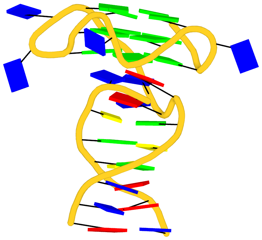
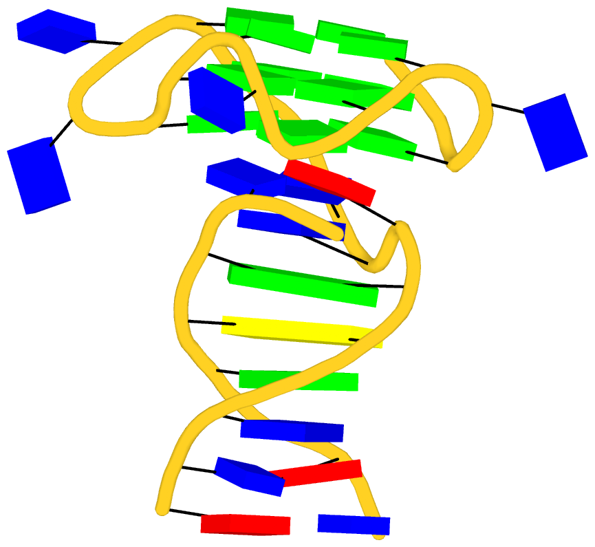
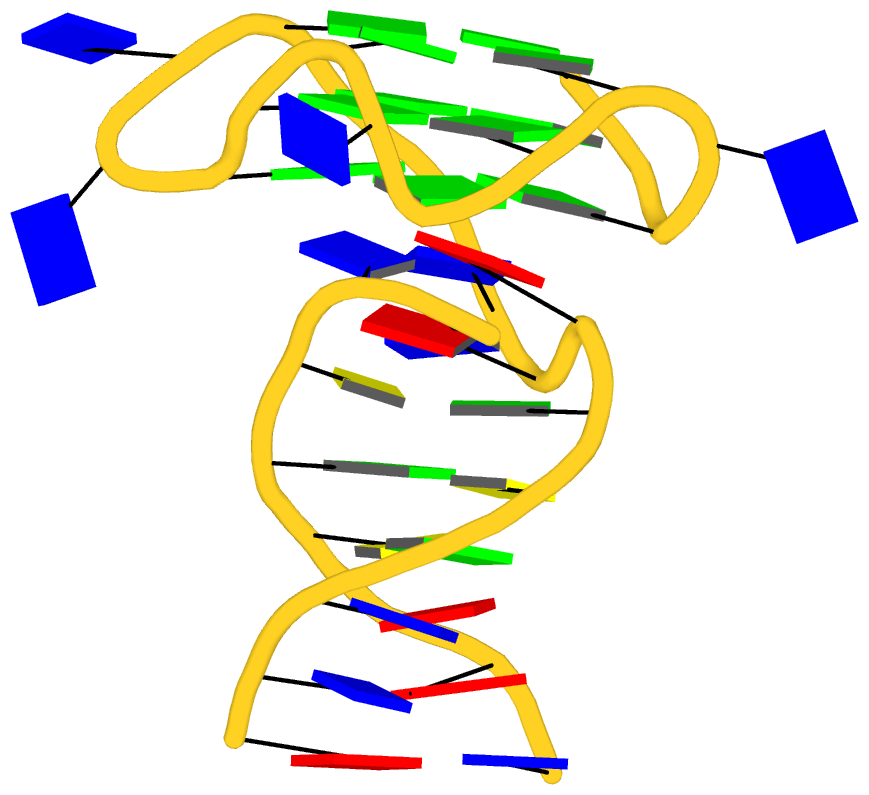
As its title suggests, however, this blog post is about the cartoon-block representations. Four styles of such schematics are shown below, which can all be easily generated using DSSR/PyMOL.
 |
 |
| in default style |
with base-pair blocks |
 |
 |
| minor-groove highlighted |
top-face highlighted |
The cartoon-block representations possess unique features not seen elsewhere. With the help of the dssr_block in PyMOL, they are extremely easy to generate. Such schematics are likely to become popular in illustrations of nucleic acid structures.

As of today (2016-01-16), the number of registrations on the 3DNA Forum has reached 2,562. Moreover, all the members (as far as I can tell) are legitimate since the Forum has remained spam free. From the very beginning, ensuring a high information-to-noice ratio has been a top priority. The goal has been achieved by taking the following measures:
State the rules clearly in the “Registration Agreement”
This forum is dedicated to topics generally related to the 3DNA suite of software programs for the analysis, rebuilding and visualization of three-dimensional nucleic acid structures. To make the 3DNA forum a more pleasant virtual community for all of us to learn from and contribute to, please be considerate and practice good netiquette (http://www.albion.com/netiquette/).
I strive to make the forum spam free. Specifically, posts that are not 3DNA related in the broad sense are taken as spams, and are strictly forbidden. You are solely responsible for the content of your posts. We reserve the right to remove any post deemed as inappropriate, deactivate the account and ban the IP address of any abuser of the forum, WITHOUT NOTICE.
When posting on the Forum, please abide by the following rules: …
In a nutshell, you are welcome to participate and should not hesitate to ask questions, but remember to play nice and preferably share what you’ve learned! Please note that we do not tolerate spamming or off-topic trolling of any form.
Take advantage of anti-spam software
In additional to the verification of email address and check for black-listed IP addresses, the topic-specific questions have been very effective. Three examples of such questions are shown below:
What does the 'A' in 3DNA stand for? (hint: 4-char long)
How many standard bases does RNA have (hint: 1-digit number)
What is the value of the expression (3.1498 * 0 + 168)?
Overall, I do not like CAPTCHA — I’ve found the highly-distorted images in some websites especially troublesome. For the first few of years (to ~2014), the 3DNA Forum did not contain a captcha image in the registration page. Later on, however, I’ve noticed quite a few spam registrations/posts. In addition to quickly cleaning them up manually, I had refined the topic-specific questions, and turned on the visual verification image at level “Medium — Overlapping colored letters, with noise/lines”. Experience over the past couple of year has demonstrated the effectiveness of the combined strategy. As shown in the screen capture below, as of this writing, 177,562 spammers have been blocked by the anti-spam software!

Verify and approve ‘suspect’ accounts quickly
The above mentioned anti-spaming measures have blocked virtually all the “bad guys” so I do not need to waste time fighting them. I receive an email notification for each successful registration. The vast majority of registrants can then immediately access the member-only download section or post questions on the 3DNA Forum after registration. A significant portion (~1 out of 6) of the registrations, however, would be masked as suspicious and need my action. The email message for such cases reads like this:
‘xxxx’ has just signed up as a new member of your forum. Click the link below to view their profile. …
Before this member can begin posting they must first have their account approved. Click the link below to go to the approval screen. …
Wherever I have access to the Internet (including after hours with an iPad Air 2), I’ve always been quick in verifying and (mostly) approving these registrations.
Overall, since http://forum.x3dna.org was created in December 2011, the Forum has received significant attention in the field of DNA/RNA structural bioinformatics. As the community begins to appreciate and fully take advantage of what DSSR and SNAP have to offer, I have no doubt the Forum will gain even wider-spread recognition.

In recent years, reproducibility of ‘scientific’ publications has become quite a topic. See a recent essay Five selfish reasons to work reproducibly by Markowetz in Genome Biology (2015, 16:274). There are numerous reasons why reproducibility could become an issue at all in science. What I have continuously strived for in my scientific career, however, is to ensure that my published results are reproducible. As a concrete example, I created a dedicated section titled DSSR-NAR paper on the 3DNA Forum that provides full details (scripts and data files) so that any interested parties can rigorously reproduce the results reported in the DSSR Nucleic Acids Research (NAR) paper.
In my support of 3DNA for over a decade, the #1 issue I experienced is undoubtedly vague (non-reproducible) questions. For example, I have recently been asked via email why the 3DNA find_pair/analyze programs miss “some basepair … even though it is in the pdb file”. Without access to the PDB file to reproduce the problem, however, I cannot provide a concrete answer. In an effect to prevent ambiguous questions, I made the following explicit point in the “Registration Agreement” of the 3DNA Forum (no. 2 on the list):
Be specific with your questions; provide a minimal, reproducible example if possible; use attachments where appropriate.
The #2 issue is receiving 3DNA-related questions privately instead of on the intended public 3DNA Forum. I turned off “personal messaging” to receive private messages on the Forum long time ago, yet I have kept receiving questions via emails. In several locations on the 3DNA Forum, I have made this ‘public-question’ policy crystal clear:
Ask your questions in the public 3DNA forum instead of sending xiangjun emails or personal messages. (no. 1 on the ‘Registration Agreement’)
Please be aware that for the benefit of the 3DNA-user community at large, I do not provide private email/personal message support; the forum has been created specifically for open discussions of all 3DNA-related issues. In other words, any 3DNA-associated questions are welcome and should be directed here. Presumably I’ve made the message simple and clear enough to get across without further explanation. (in ‘Site announcements » Download instructions’ and ‘Downloads » 3DNA download’)
In response to the many 3DNA-related questions that still keep coming via email, I created the following entry of Canned Responses in gmail:
Thanks for your interest in using 3DNA. Please be aware that for the benefit of the 3DNA-user community at large, I do not provide private email support; the 3DNA Forum (http://forum.x3dna.org/) has been created specifically for open discussions of all 3DNA-related issues. In other words, any 3DNA-associated questions are welcome and should be directed there. I monitor the forum regularly and respond to posts promptly.
I look forward to seeing you on the 3DNA Forum (http://forum.x3dna.org/).
Overall, I’ve learned from experience that addressing reproducible questions publicly does the best for the 3DNA community. Users can register with personal (free) email address, and post simulated data to illustrate the problem at hand. Moreover, questions on the Forum have always received quick responses. Over time, the Forum has served as an archive that everyone can benefit from.

As of v2.3-2016jan01, the 3DNA analyze program outputs a list of new ‘simple’ base-pair and step parameters, by default. Shown below is a sample output for PDB entry 1xvk. This echinomycin-(GCGTACGC)2 complex has a single DNA strand as the asymmetric unit. 3DNA needs the the biological unit (1xvk.pdb1) to analyze the duplex (with the -symm option). This structure contains two Hoogsteen base pairs, and has popped up on the 3DNA Forum for the zero or negative Rise values. Note that the ‘simple’ Rise values are all positive; for the middle (#4) TA/TA step, it is now 3.09 Å instead of 0.
# find_pair -symm 1xvk.pdb1 1xvk.bps
# analyze -symm 1xvk.bps
#   OR by combing the above two commands:
# find_pair -symm 1xvk.pdb1 | analyze -symm
# The output is in file '1xvk.out'
This structure contains 4 non-Watson-Crick (with leading *) base pair(s)
----------------------------------------------------------------------------
Simple base-pair parameters based on RC8--YC6 vectors
      bp        Shear    Stretch   Stagger    Buckle  Propeller  Opening
*    1 G+C      -3.07      1.55     -0.35     -6.98      0.29     67.33
     2 C-G       0.27     -0.17      0.35    -22.34      3.33     -2.80
     3 G-C      -0.39     -0.17      0.41     22.91      1.81     -2.73
*    4 T+A      -3.29      1.56      0.31     -8.03      1.59    -70.46
*    5 A+T      -3.29      1.56     -0.31     -8.03      1.59     70.46
     6 C-G       0.39     -0.17      0.41    -22.91      1.81     -2.72
     7 G-C      -0.27     -0.17      0.35     22.34      3.32     -2.80
*    8 C+G      -3.07      1.55      0.35     -6.98      0.30    -67.33
           ~~~~~~~~~~~~~~~~~~~~~~~~~~~~~~~~~~~~~~~~~~~~~~~~~~~~~~~~~~~~
       ave.     -1.59      0.69      0.19     -3.75      1.75     -1.38
       s.d.      1.72      0.92      0.32     17.57      1.15     52.11
----------------------------------------------------------------------------
Simple base-pair step parameters based on consecutive C1'-C1' vectors
      step       Shift     Slide      Rise      Tilt      Roll     Twist
*    1 GC/GC     -0.55      0.39      7.41      6.40     -4.22     23.36
     2 CG/CG     -0.05      0.87      2.44     -0.55      3.94     -0.81
*    3 GT/AC      0.38      0.47      7.23     -8.62      3.75     25.70
*    4 TA/TA     -0.00      4.73      3.09     -0.00      7.49     25.67
*    5 AC/GT     -0.38      0.47      7.23      8.62      3.75     25.70
     6 CG/CG      0.05      0.87      2.44      0.55      3.94     -0.82
*    7 GC/GC      0.55      0.39      7.41     -6.40     -4.22     23.36
            ~~~~~~~~~~~~~~~~~~~~~~~~~~~~~~~~~~~~~~~~~~~~~~~~~~~~~~~~~~~~
        ave.     -0.00      1.17      5.32     -0.00      2.06     17.45
        s.d.      0.39      1.59      2.50      6.21      4.49     12.52
The simple parameters are ‘intuitive’ for non-Watson-Crick base pairs and associated base-pair steps, where the existing standard-reference-frame-based 3DNA parameters may look weird. Note that these simple parameters are for structural description only, not to be fed into the ‘rebuild’ program. Overall, they complement the rigorous characterization of base-pair geometry, as demonstrated by the original analyze/rebuild pair of programs in 3DNA.
In short, the ‘simple’ base-pair parameters employ the YC6—RC8 vector as the y-axis whereas the ‘simple’ step parameters use consecutive C1’—C1’ vectors. As before, the z-axis is the average of two base normals, taking consideration of the M–N vs M+N base-pair classification. In essence, the ‘simple’ parameters make geometrical sense by introducing an ad hoc base-pair reference frame in each case. More details will be provided in a series of blog posts shortly.
Overall, this new section of ‘simple’ parameters should be taken as experimental. The output can be turned off by specifying the analyze -simple=false command-line option explicitly. As always, I greatly appreciate your feedback.

Over the years, I have played quite a few computer programming languages. ANSI C has become my top choice for ‘serious’ software projects, due to its small size, efficiency, flexibility, and ubiquitous support. Moreover, C is a mature language, with a rich ecosystem. As it turns out, C has also been consistently rated as one of the most popular computer languages (#1 or #2) over the past thirty years.
Needless to say, ANSI C has its own quirks, and it takes a steep learning curve. However, once you get over the hurdles, the language serves you. I cannot remember when, but it has been a long while that coding in ANSI C is no longer an issue. It is the understanding of scientific questions that takes most of my time, and coding helps greatly in refining my thoughts.
Not surprisingly, ANSI C was chosen as the sole language for DSSR (and SNAP, or 3DNA in general). The ensure the overall quality of the DSSR codebase, I have taken the following steps:
- The whole project is under git.
- The ANSI C source code is compiled with strict GCC options for full compliance to the standard:
-ansi -pedantic -W -Wall -Wextra -Wunused -Wshadow -Werror -O3
- The executable is checked with valgrind for any memory leak:
valgrind --leak-check=full x3dna-dssr -i=1ehz.pdb -o=1ehz.out --quiet
==19624== Memcheck, a memory error detector
==19624== Copyright (C) 2002-2013, and GNU GPL'd, by Julian Seward et al.
==19624== Using Valgrind-3.10.1 and LibVEX; rerun with -h for copyright info
==19624== Command: x3dna-dssr -i=1ehz.pdb -o=1ehz.out --quiet
==19624==
==19624==
==19624== HEAP SUMMARY:
==19624==     in use at exit: 0 bytes in 0 blocks
==19624==   total heap usage: 52,829 allocs, 52,829 frees, 92,878,578 bytes allocated
==19624==
==19624== All heap blocks were freed -- no leaks are possible
==19624==
==19624== For counts of detected and suppressed errors, rerun with: -v
==19624== ERROR SUMMARY: 0 errors from 0 contexts (suppressed: 0 from 0)
- Extensive tests (with the simple
diff command) to ensure the program is working as expected.
The above four measures combined allow me to add new features, refactor the code, and fix bugs, without worrying about accidentally breaking existing functionality. Reading literature (including citations to 3DNA/DSSR) and responding to user feedback on the 3DNA Forum keep me continuously improve DSSR. Some of the recent refinements to DSSR came about this way.


